. 2018 Jun;15(6):618-629.
doi: 10.1038/cmi.2017.5. Epub 2017 Mar 13.

Glucocorticoid receptor promotes the function of myeloid-derived suppressor cells by suppressing HIF1α-dependent glycolysis

Affiliations

Glucocorticoid receptor promotes the function of myeloid-derived suppressor cells by suppressing HIF1α-dependent glycolysis

Yun Lu et al. Cell Mol Immunol. 2018 Jun.

Abstract

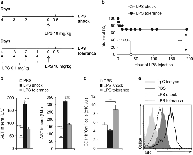
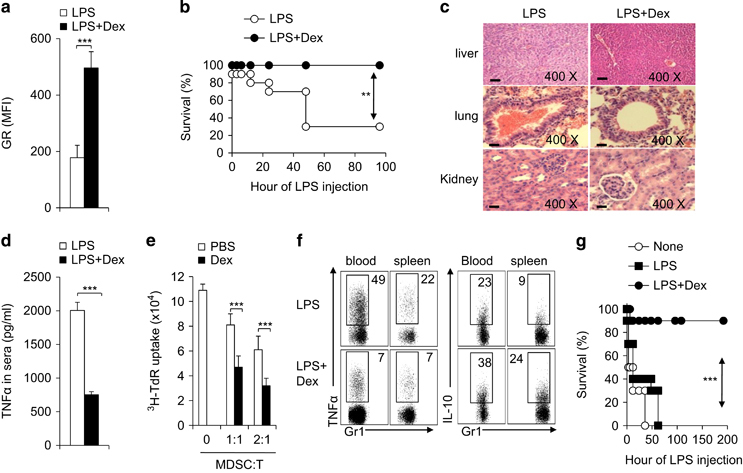
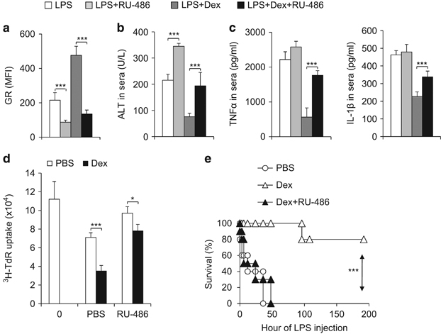
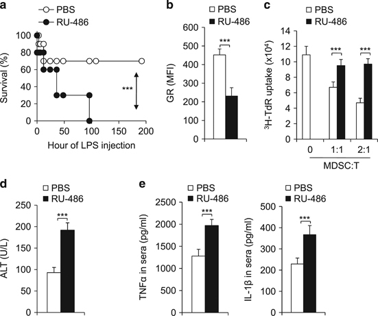
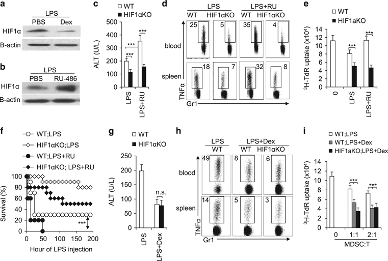
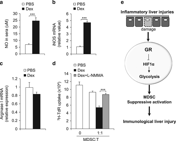
Immunomodulatory signaling imposes tight regulations on metabolic programs within immune cells and consequentially determines immune response outcomes. Although the glucocorticoid receptor (GR) has been recently implicated in regulating the function of myeloid-derived suppressor cells (MDSCs), whether the dysregulation of GR in MDSCs is involved in immune-mediated hepatic diseases and how GR regulates the function of MDSCs in such a context remains unknown. Here, we revealed the dysregulation of GR expression in MDSCs during innate immunological hepatic injury (IMH) and found that GR regulates the function of MDSCs through modulating HIF1α-dependent glycolysis. Pharmacological modulation of GR by its agonist (dexamethasone, Dex) protects IMH mice against inflammatory injury. Mechanistically, GR signaling suppresses HIF1α and HIF1α-dependent glycolysis in MDSCs and thus promotes the immune suppressive activity of MDSCs. Our studies reveal a role of GR-HIF1α in regulating the metabolism and function of MDSCs and further implicate MDSC GR signaling as a potential therapeutic target in hepatic diseases that are driven by innate immune cell-mediated systemic inflammation.


Conflict of interest statement

The authors declare no conflict of interest.

Figures

Figure 1
Figure 1
The alternation of myeloid-derived suppressor cell (MDSC) glucocorticoid-receptor in immune-mediated hepatic injuries. (a) diagram of the experimental model for LPS tolerance and for LPS shock induction; the mouse survival curve is plotted in b. In the indicated murine models, the serum ALT and AST levels were determined with ELISA (c); the blood CD11b+Gr1+ cells were analysed by FACS (d); FACS analysis of GR expression in CD11b+Gr1+ cells (e). Data are representative of three independent experiments (n=4–10). **P<0.01 and ***P<0.001 compared with the indicated groups. GR, glucocorticoid receptor; LPS, lipopolysaccharide.
Figure 2
Figure 2
Dex treatment upregulates GR expression, potentiates MDSC activities and protects mice against immune-hepatic injuries. Age-matched C57BL/6 mice were injected i.p. with PBS (solvent) or Dex (5 mg/kg body weight) daily starting at 6 h before LPS (5 mg/kg) injection. The GR expression on the CD11b+Gr1+ cells from liver 72 h following LPS injection was determined by FACS (a) and the survival curve of experimental mice is plotted (b, n=10). In a separate set of animals, the mice were sacrificed at 72 h following LPS-injection. The pathological changes in mouse liver, lung and kidney were examined by histochemistry (c). TNFα level in serum was determined with ELISA (d). The immunosuppressive activity of myeloid-derived CD11b+Gr1+ cells was determined by mixed lymphocyte reaction (e). The TNFα and IL-10 production in CD11b+Gr1+ cells in blood and spleen was analysed by FACS (f). Three groups of donor C57BL/6 mice were challenged with PBS (None), LPS or LPS plus Dex. After 3 days, a total of 1x10⁶ CD11b+Gr1+ cells were sorted from the liver of indicated mice and adoptively transferred into C57BL/6 recipient mice (10 mice per group) via i.v. Injection. After 10-12 h, all groups of recipient mice were challenged with LPS (10 mg/kg) and mouse survival was followed (g). Data are representative of three (ce) or four (f) independent experiments (n=3–10). **P<0.01 and ***P<0.001 compared with the indicated groups. GR, glucocorticoid receptor; IL-10, interleukin-10; i.v., intravenous; LPS, lipopolysaccharide; MDSC, myeloid-derived suppressor cell; PBS, phosphate-buffered saline; TNFα, tumor necrosis factor-α.
Figure 3
Figure 3
The downregulation of GR expression by RU-486 counteracts the effect of Dex in protecting mice against immune-hepatic injuries. As described in Figure 2, IMH was induced in four groups of age-matched C57BL/6 mice with the indicated treatments. Then, 72 h after LPS challenge, GR expression of CD11b+Gr1+MDSC in liver was analysed by FACS (a) and the level of ALT (b) TNFα and IL-1β (c) in serum were determined by ELISA. The immunosuppressive activity of myeloid-derived CD11b+Gr1+ cells was determined by mixed lymphocyte reaction (d) Three groups of donor C57BL/6 mice were challenged with LPS, LPS plus Dex or LPS plus Dex and RU-486 (10 mg/kg, i.p.). After 3 days, a total of 1 × 10⁶ CD11b+Gr1+ cells were sorted from the liver of indicated mice and adoptively transferred into C57BL/6 recipient mice (10 mice per group) via i.v. injection. After 10–12 h, all groups of recipient mice were challenged with LPS (10 mg/kg), and mouse survival was followed (e) Data are representative of three (ac) or two (d) independent experiments (ad; n=3–5). ***P<0.001 compared with the indicated groups. GR, glucocorticoid receptor; IMH, immunological hepatic injury; i.p., intraperitoneal; MDSC, myeloid-derived suppressor cell; LPS, lipopolysaccharide.
Figure 4
Figure 4
The downregulation of GR expression by RU-486 abolishes immune tolerance in an LPS-mediated tolerance model. Two groups of aged-matched C57BL/6 mice (10 per group) were pretreated with low dose LPS (0.1 mg/kg) daily from day 4 prior to challenge with LPS (10 mg/kg) for LPS tolerance induction. Either PBS or RU-486 was administrated at 6 h before the last LPS challenge. Mouse survival curve is plotted (a). Then, 72 h after the LPS challenge, GR expression of CD11b+Gr1+MDSC in liver was analysed by FACS (b) the immunosuppressive activity of myeloid-derived CD11b+Gr1+ cells was determined by mixed lymphocyte reaction (c) and the levels of ALT (d) TNFα and IL-1β (e) in serum were determined by ELISA. Data are representative of three (be) independent experiments (n=3–5). ***P<0.001 compared with the indicated groups. GR, glucocorticoid receptor; IL-1β, interleukin-1β; LPS, lipopolysaccharide; PBS, phosphate-buffered saline; TNFα, tumor necrosis factor-α.
Figure 5
Figure 5
Blocking glycolysis protects mice against immune-hepatic injuries in IMH. As described in Figure 2, IMH was induced in the indicated three groups of age-matched C57BL/6 mice. Then, 12 h after LPS challenge, CD11b+Gr1+ cells were sorted from livers of indicated mice and were stimulated with LPS or LPS+IFN-γ for 10 h. The glycolytic activity (a) or the mRNA expression of indicated glycolytic genes (b) in the indicated groups was measured by the generation of 3H-labeled H2O from [3-3H]-glucose or by qPCR. The liver MDSCs were isolated from IMH mice and were stimulated by LPS for 10 h in the presence of vehicle (PBS) or 1 mmol/l 2-DG. The glycolytic activity was measured as described above (c). As described in Figure 2, IMH was induced in three groups of age-matched C57BL/6 mice with the indicated treatments (LPS 10 mg/kg). The survival curve is plotted (d) Then, 72 h after the LPS challenge, the immunosuppressive activity of myeloid-derived CD11b+Gr1+ cells was determined by mixed lymphocytes reaction (e) and the levels of ALT (f) and TNFα (g) in serum were determined by ELISA. Data are representative of two (ac) or three (eg) independent experiments (n=3–10). *P<0.05, **P<0.01 and ***P<0.001 compared with the indicated groups. IMH, immunological hepatic injury; IFN-γ, interferon-γ; LPS, lipopolysaccharide; MDSC, myeloid-derived suppressor cell; qPCR, quantitative PCR; TNFα, tumor necrosis factor-α.
Figure 6
Figure 6
HIF1α is a downstream effector of GR signaling in MDSCs in IMH. The protein level of HIF1α in liver MDSCs that were isolated from IMH mice with the indicated treatments was determined by western blot (a, b). As described in Figure 2, IMH was induced in WT or HIF1αfl/fl, LysM Cre+ mice with the indicated treatments. Then, 72 h after LPS-injection, the ALT level in serum was determined with ELISA (c, g) the TNFα production in CD11b+Gr1+ cells in blood and spleen was analysed by FACS (d, h) and the immunosuppressive activity of myeloid-derived CD11b+Gr1+ cells was determined by mixed lymphocyte reaction (e, i) As described in Figure 2, IMH was induced in age-matched WT or HIF1αfl/fl, LysM Cre+ mice (n=10) with the indicated treatments (LPS 5 mg/kg). The survival curve is plotted (f). Data are representative of two (ae) or three (gi) independent experiments (n=3–5). ***P<0.001 compared with the indicated groups. GR, glucocorticoid receptor; IMH, immunological hepatic injury; MDSC, myeloid-derived suppressor cell; LPS, lipopolysaccharide; NS, not significant; TNFα, tumor necrosis factor-α; WT, wild type.
Figure 7
Figure 7
GR signaling modulates NO production in CD11b+Gr1+ MDSCs in IMH. As described in Figure 2, IMH was induced in mice with the indicated treatments. Then, 72 h after LPS-injection, the NO level in serum was determined by Greiss assay (a), the mRNA level of indicated genes in CD11b+Gr1+ cells was determined by qPCR (b,c) and the immunosuppressive activity of myeloid-derived CD11b+Gr1+ cells was determined by mixed lymphocyte reaction (d). Data are representative of three independent experiments (n=4–5). ***P<0.001 compared with the indicated groups. (e) A model summarizes the role of GR signaling in establishing MDSC functions through HIF1α dependent glycolytic pathway. GR, glucocorticoid receptor; IMH, immunological hepatic injury; MDSC, myeloid-derived suppressor cell; qPCR, quantitative PCR.

References

    1. Bowen DG, Walker CM. Adaptive immune responses in acute and chronic hepatitis C virus infection. Nature. 2005;436:946–952. doi: 10.1038/nature04079. - DOI - PubMed
    1. Das M, Sabio G, Jiang F, Rincon M, Flavell RA, Davis RJ. Induction of hepatitis by JNK-mediated expression of TNF-alpha. Cell. 2009;136:249–260. doi: 10.1016/j.cell.2008.11.017. - DOI - PMC - PubMed
    1. Liberal R, Grant CR, Longhi MS, Mieli-Vergani G, Vergani D. Diagnostic criteria of autoimmune hepatitis. Autoimmun Rev. 2014;13:435–440. doi: 10.1016/j.autrev.2013.11.009. - DOI - PubMed
    1. Yang H, Bi Y, Xue L, Wang J, Lu Y, Zhang Z, et al. Multifaceted Modulation of SIRT1 in Cancer and Inflammation. Crit Rev Oncog. 2015;20:49–64. doi: 10.1615/CritRevOncog.2014012374. - DOI - PubMed
    1. Zhao Y, Wu T, Shao S, Shi B. Phenotype, development, and biological function of myeloid-derived suppressor cells. Oncoimmunology. 2016;5:e1004983. doi: 10.1080/2162402X.2015.1004983. - DOI - PMC - PubMed