. 2017 Mar 13:7:44189.
doi: 10.1038/srep44189.

Endogenous TNFα orchestrates the trafficking of neutrophils into and within lymphatic vessels during acute inflammation

Affiliations

Endogenous TNFα orchestrates the trafficking of neutrophils into and within lymphatic vessels during acute inflammation

Samantha Arokiasamy et al. Sci Rep. .

Abstract

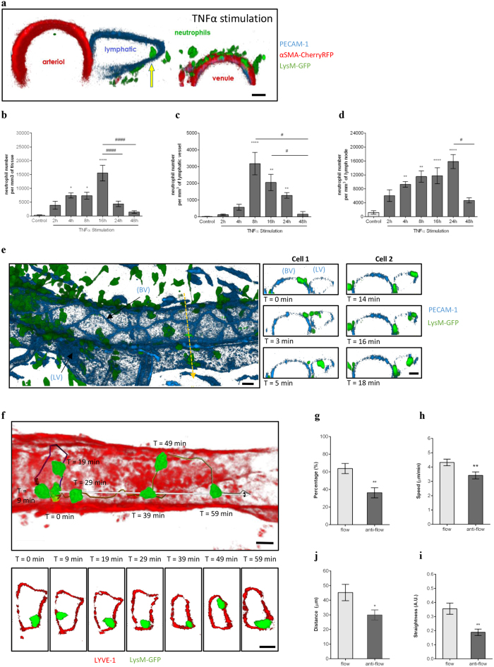
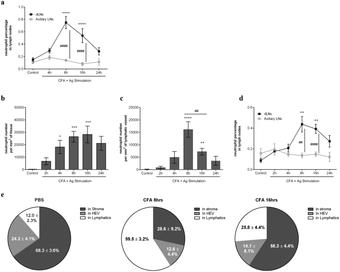
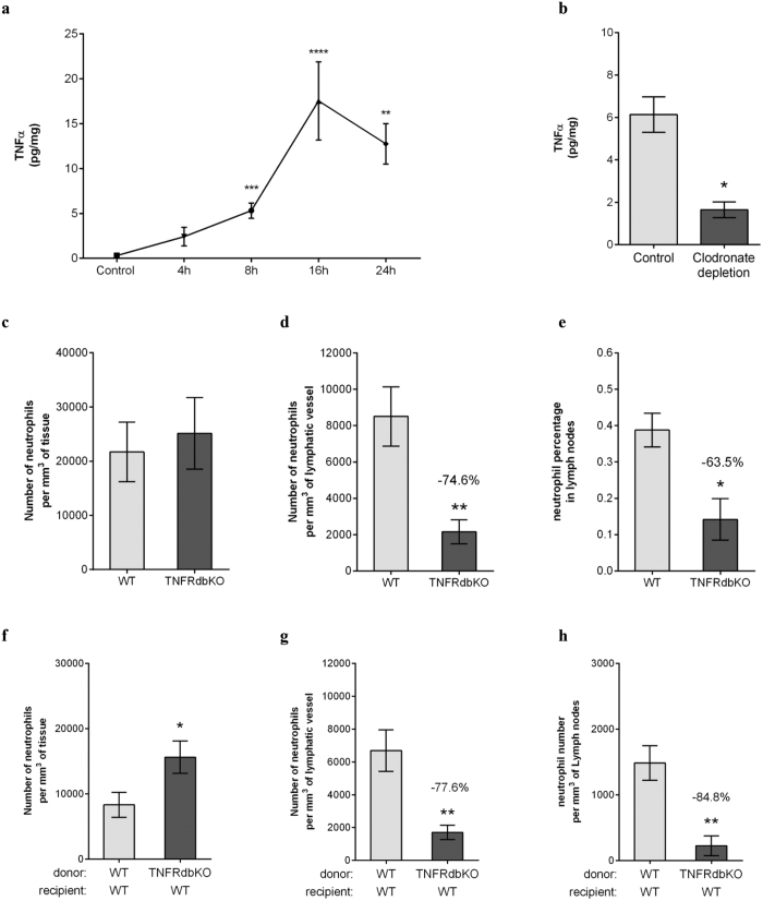
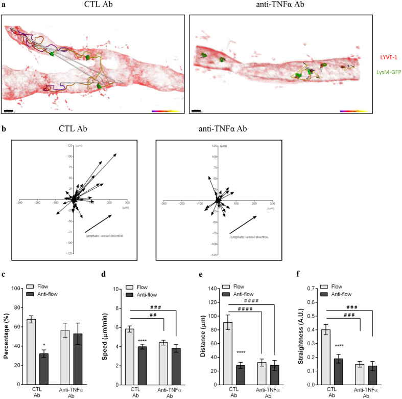
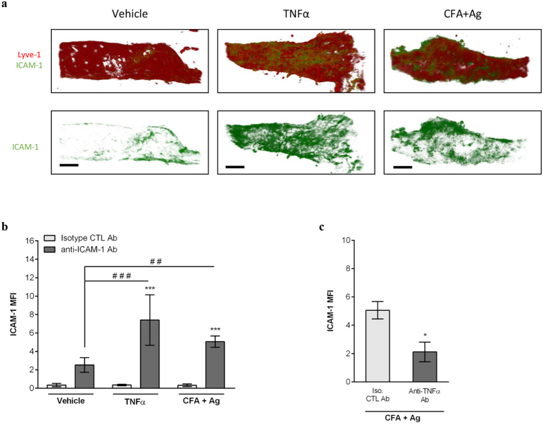
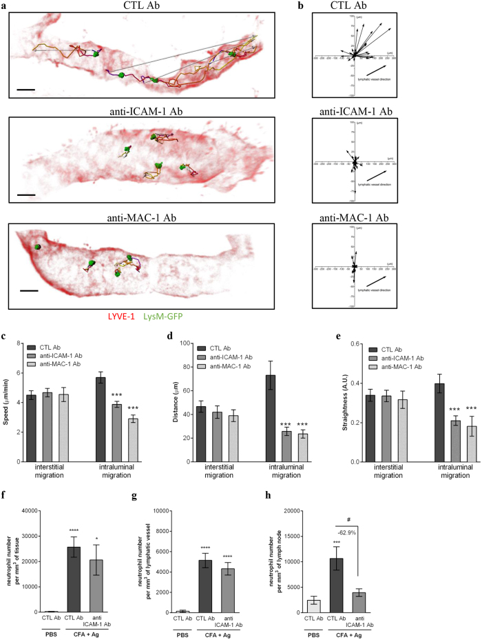
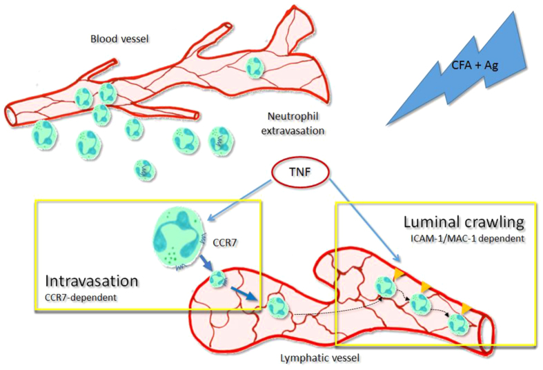
Neutrophils are recognised to play a pivotal role at the interface between innate and acquired immunities following their recruitment to inflamed tissues and lymphoid organs. While neutrophil trafficking through blood vessels has been extensively studied, the molecular mechanisms regulating their migration into the lymphatic system are still poorly understood. Here, we have analysed neutrophil-lymphatic vessel interactions in real time and in vivo using intravital confocal microscopy applied to inflamed cremaster muscles. We show that antigen sensitisation of the tissues induces a rapid but transient entry of tissue-infiltrated neutrophils into lymphatic vessels and subsequent crawling along the luminal side of the lymphatic endothelium. Interestingly, using mice deficient in both TNF receptors p55 and p75, chimeric animals and anti-TNFα antibody blockade we demonstrate that tissue-release of TNFα governs both neutrophil migration through the lymphatic endothelium and luminal crawling. Mechanistically, we show that TNFα primes directly the neutrophils to enter the lymphatic vessels in a strictly CCR7-dependent manner; and induces ICAM-1 up-regulation on lymphatic vessels, allowing neutrophils to crawl along the lumen of the lymphatic endothelium in an ICAM-1/MAC-1-dependent manner. Collectively, our findings demonstrate a new role for TNFα as a key regulator of neutrophil trafficking into and within lymphatic system in vivo.


Conflict of interest statement

The authors declare no competing financial interests.

Figures

Figure 1
Figure 1. Dynamics of neutrophil migration into cremaster muscle lymphatics upon TNFα-stimulation.
The dynamics of neutrophil migration into the tissue and lymphatic vessels was analysed by intravital confocal microscopy in TNFα-stimulated mouse cremaster muscles. (a) Representative 3D-reconstructed still image (2 μm cross-section) from a LysM-GFP × αSMA-CherryRFP mouse [exhibiting both endogenous GFP-fluorescent neutrophils (green) and RFP-fluorescent pericytes/smooth muscle cells (red) and immunostained with a non-blocking anti-PECAM-1 mAb (blue)] cremaster tissue showing a neutrophil within the lymphatic vessel (yellow arrow) post TNFα-stimulation. (b) Time-course of neutrophil extravasation in TNFα-stimulated cremaster muscles. (c) Time-course of neutrophil migration into lymphatic vessels upon TNFα-stimulation. (d) Total neutrophil-infiltrate in dLNs upon TNFα-stimulation. (e) Representative 3D-reconstructed still image of a post-capillary venule and an adjacent lymphatic vessel from a LysM-GFP mouse (immunostained with non-blocking anti-PECAM-1 mAb (blue)]. The right panel images illustrate a time-lapse series of 2 μm-thick cross-sections along the z-plane (dotted-yellow arrow) showing the migration of two neutrophils (Cell-1 & Cell-2) into the lymphatic vessel. (f) Representative 3D-reconstructed still image of a lymphatic vessel from a TNFα-stimulated cremaster tissue of a LysM-GFP mouse and immunostained with an anti-LYVE-1 mAb (red) in vivo. Neutrophil crawling path (colour-coded line) and directionality (white arrow) is shown on the image. The bottom panel images are a series of high magnification cross-sections of the main image at indicated time-points illustrating the continuous attachment of the neutrophil to the lymphatic endothelium. (g) Percentage of neutrophils crawling in the afferent direction (flow) or in the opposite direction (anti-flow). Speed (h), directionality (i) and straightness (j) of neutrophils crawling in the afferent (flow) or opposite direction (anti-flow) of the cremaster lymphatic vessels. Data are expressed as mean ± SEM from 5–12 animals per group (at least 5 independent experiments). For the crawling parameter analysis, a total of 63 cells were quantified from 8 mice. Statistically significant differences between stimulated and unstimulated treatment groups are indicated by asterisks: *P < 0.05; **P < 0.01; ***P < 0.001; ****P < 0.0001. Significant differences between responses at different time points are indicated by hash symbols: #P < 0.05; ####P < 0.0001. Bar = 10 μm.
Figure 2
Figure 2. Neutrophil rapidly migrates into lymphatic system of the cremaster muscle during antigen sensitisation in vivo.
Neutrophil migration into the lymphatic system was induced in WT animals following antigen sensitisation with complete Freund’s adjuvant (CFA+Ag). (a) Time course of neutrophil migration into draining LNs (inguinal) or non-draining LNs (axillary) of mice injected intradermally with CFA+Ag and as analysed by flow cytometry. (b) Time course of CFA+Ag-induced neutrophil extravasation in mice injected intra-scrotally with CFA+Ag as visualised by confocal microscopy. (c) Time course of CFA+Ag-induced neutrophil intravasation into the cremaster lymphatic vessels of WT mice as visualised by confocal microscopy. (d) Time course of neutrophil migration into draining and non-draining LNs in mice as analysed by flow cytometry. (e) Quantification of neutrophil localisation in the dLNs of mice stimulated intra-scrotally with CFA (8 or 16 hrs) or with PBS (control), and as analysed by confocal microscopy. Data are represented as percentages of neutrophils present in the HEV, LYVE-1+ vessels and in the stroma of the LNs. Data are expressed as mean ± SEM of N = 5–12 animals (~10 images per cremasters for confocal microscopy) per group from at least 5–10 experiments. Statistically significant differences between stimulated and control groups or between WT and TNFRdbKO mice are indicated by asterisks: *P < 0.05; **P < 0.01; ***P < 0.001; ****P < 0.0001. Significant differences between other groups are indicated by hash symbols: ##P < 0.01; ####P < 0.0001.
Figure 3
Figure 3. TNFα instruct the neutrophils to migrate into the lymphatic system upon antigen sensitisation.
Neutrophil migration into the lymphatic system of the cremaster muscle following antigen sensitisation with complete Freund’s adjuvant (CFA+Ag) was induced in WT and TNFRdbKO animals as well as in chimeric animals exhibiting neutrophils deficient in TNFRs. (a) Time course of TNFα release in the cremaster muscles of WT mice following intra-scrotal injection of CFA+Ag and as quantified by ELISA. (b) TNFα release in mice subjected to clodronate liposome-induced macrophage depletion. (c) Number of extravasated neutrophils in cremaster muscles of WT and TNFRdbKO mice at 16 hrs post-CFA+Ag-stimulation as quantified by confocal microscopy. (d) Number of neutrophils within cremaster lymphatic vessels of WT and TNFRdbKO mice at 16 hrs post-CFA+Ag-stimulation as quantified by confocal microscopy. (e) Percentage of neutrophils in dLNs of WT and TNFRdbKO mice at 16 hrs post-CFA+Ag-stimulation as quantified by flow cytometry. (f) Number of extravasated neutrophils in cremaster muscles at 16 hrs post-CFA+Ag-stimulation from chimeric animals receiving bone marrow transplant from WT or TNFRdbKO donor mice and as quantified by confocal microscopy. (g) Number of neutrophils within cremaster lymphatic vessels at 16 hrs post-CFA+Ag-stimulation from chimeric animals receiving bone marrow transplant from WT or TNFRdbKO donor mice and as quantified by confocal microscopy. (h) Number of neutrophils found in the dLNs of chimeric animals receiving bone marrow transplant from WT or TNFRdbKO donor mice as quantified by confocal microscopy 16 hrs post-CFA+Ag-stimulation. Data are expressed as mean ± SEM of N = 5–12 animals per group from at least 5–10 experiments. Statistically significant differences between stimulated and control groups or between WT and TNFRdbKO mice are indicated by asterisks: *P < 0.05; **P < 0.01; ****P < 0.0001.
Figure 4
Figure 4. TNFα promotes CCR7-dependent migration of neutrophils into lymphatic vessels in vivo.
(a) Analysis by flow cytometry of CCR7 expression (intracellular and cell-surface) on neutrophils isolated from the blood circulation, CFA+Ag-stimulated-cremaster muscles and dLNs of WT and CCR7KO animals. (b) CCR7 surface expression on tissue-infiltrated neutrophils from WT and TNFRdbKO mice subjected to CFA+Ag-induced inflammation. (c–e) WT and CCR7KO mice were subjected to TNFα-induced cremaster muscle inflammation and neutrophil responses in the tissue and dLNs was assessed by confocal microscopy 16 hrs post-inflammation. (c) Number of extravasated neutrophils in of WT and CCR7KO mice. (d) Number of intravasated neutrophils in lymphatic vessels of cremaster muscles from WT and CCR7KO mice. (e) Neutrophil number in the cremaster dLNs of WT and CCR7KO animals. (f–k) WT, CCR7KO mice or CCR7KO-neutrophil chimeric animals were subjected to antigen sensitisation (CFA+Ag) and neutrophil responses in the cremaster muscle and dLNs (16 hrs post-inflammation) was assessed by confocal microscopy. (f) Number of extravasated neutrophils in inflamed cremaster muscles of WT and CCR7KO mice. (g) Number of intravasated neutrophils in cremaster lymphatic vessels of WT and CCR7KO mice. (h) Neutrophil number in the cremaster dLNs of WT and CCR7KO mice. (i) Number of neutrophils recruited to the cremaster muscles from lethally irradiated WT animals receiving bone marrow transplant from either WT or CCR7KO donor mice. (j) Number of neutrophils within cremaster lymphatic vessels post-CFA+Ag-stimulation from lethally irradiated WT animals receiving bone marrow transplant from either WT or CCR7KO donor mice. (k) Neutrophil number in the cremaster dLNs from lethally irradiated WT animals receiving bone marrow transplant from either WT or CCR7KO donor mice. Data are expressed as mean ± SEM of N = 7–12 animals per group (from at least 5 independent experiments). Statistically significant differences between stimulated/specific mAb and unstimulated treatment/isotype control groups are indicated by asterisks: *P < 0.05; **P < 0.01; ***P < 0.001; ****P < 0.0001. Significant differences between responses in WT vs. CCR7KO animals (or between different tissues) are indicated by hash symbols: #P < 0.05; ##P < 0.01; ###P < 0.001; ####P < 0.0001.
Figure 5
Figure 5. TNFα controls the crawling of neutrophils into the lymphatic vessels in vivo.
The effect of anti- TNFα blocking mAb on neutrophil crawling along the luminal side of the lymphatic endothelium was analysed by intravital confocal microscopy (IVM) using LysM-GFP mice subjected to CFA+Ag-induced cremaster inflammation and immunostained in vivo with a non-blocking dose of Alexa555-conjugated anti-LYVE-1 mAb. Isotype control or anti- TNFα blocking mAbs were injected i.s. 4 hrs post-inflammation. (a) The pictures are representative still images at one time point of the IVM recording showing lymphatic-infiltrated neutrophils (green) and their associated crawling path (time-coloured mapped line) and/or directionality (arrow) as analysed by IMARIS software (LYVE-1 with an opacity filter of 5% to see the intravasated leukocytes) from CTL (left panel) or anti- TNFα (right panel) mAb-treated groups. (b) The graphs show the crawling paths of lymphatic-infiltrated neutrophils in the X & Y planes of the lymphatic vessels from CTL mAb (left panel) and anti- TNFα mAb (right panel) treated groups. (c) Quantification (in percentage) of neutrophils crawling in the afferent (flow) or opposite direction (anti-flow) of the lymphatic vessel. Mean speed (d), directionality (e) and straightness (f) of neutrophils crawling in CTL mAb and anti- TNFα treated groups. A total of 280 cells were analysed. Results are expressed as mean ± SEM of N = 4–9 mice (each mouse representing one independent experiment). Significant differences between flow and anti-flow crawling cells are indicated by *P < 0.05; ****P < 0.0001. Significant differences between the CTL and anti– TNFα mAb treated groups are indicated by hash symbols: ##P < 0.01; ###P < 0.001; ####P < 0.0001. Bar = 20 μm.
Figure 6
Figure 6. TNFα controls ICAM-1 expression on lymphatic endothelial cells in vivo.
Cremaster muscles of WT mice were stimulated with TNFα or CFA+Ag (6–8 hrs) and immunostained with Alexa555-conjugated anti-LYVE-1 and Alexa488-conjugated anti-ICAM-1 (or an isotype control) mAbs to label the lymphatic vasculature and ICAM-1, respectively. (a) The pictures are representative confocal images of cremaster lymphatic vessels showing the expression of ICAM-1 on selected lymphatic vessels from a PBS-treated control (left panels), TNFα-stimulated (middle panels) and CFA+Ag-stimulated (right panels) animals. (b) ICAM-1 expression (mean fluorescent intensity or MFI) on vessels from PBS-treated control, TNFα-stimulated and CFA+Ag-stimulated cremaster muscles as quantified by IMARIS software. (c) ICAM-1 expression on lymphatic vessels of CFA+Ag-stimulated cremaster muscles from animals pre-treated with an anti- TNFα blocking mAb or isotype CTL mAb injected 4 hrs post-inflammation. Data are expressed as mean ± SEM of N = 8–12 vessels/animals from 4 animals per group (3 independent experiments). Statistically significant differences between the staining of isotype control and anti-ICAM-1 Abs treated groups are indicated by asterisks: *P < 0.05; ****P < 0.0001. Significant differences between unstimulated and inflamed groups are indicated by hash symbols: ##P < 0.01; ####P < 0.0001. Bar = 50 μm.Results are expressed as mean ± SEM of N = 4–9 mice (1–2 vessels analysed/mouse for intravital confocal microscopy, each mice is a single experiment). Significant differences between blocking antibodies treated and control groups are indicated by *P < 0.05; ***P < 0.001; ****P < 0.0001. Significant differences between other groups are indicated by #(P < 0.05). Bar = 30 μm.
Figure 7
Figure 7. Neutrophil crawling along the lymphatic endothelium is ICAM-1/MAC-1 dependent.
LysM-GFP mice were subjected to CFA+Ag-induced cremaster inflammation for 6 hrs. Mice also received at 4.5 hrs post inflammation an i.s. injection of non-blocking dose of Alexa555-conjugated anti-LYVE-1 (red) and Alexa647-conjugated anti-PECAM-1 mAbs (not shown on the image) for the visualisation of both the lymphatic and blood vasculatures. Ninety minutes later, tissues were exteriorised to perform time-lapse recordings of the neutrophil responses for 2 hrs by intravital confocal microscopy. The effect of blocking antibodies against ICAM-1 and MAC-1 (injected locally 90 min before recordings) on neutrophil migration paths in the interstitium and lymphatic vessels was investigated and analysed using IMARIS software. (a) The pictures are representative 3D still images showing neutrophils (green) within the lymphatic vessels (red) and their respective crawling path (time-coloured mapped line) and directionality (arrow) from mice pre-treated with an isotypic control (CTL, top panel), anti-ICAM-1 (middle panel) or anti-MAC-1 (bottom panel) mAbs. (b) The graphs show the crawling paths of neutrophils in the X & Y planes of the lymphatic vessels from CTL Ab, anti-ICAM-1 and anti-MAC-1 mAbs treated groups. (c-e) The effect of anti–ICAM-1 and anti-MAC-1 blocking antibodies on neutrophil migration parameters (i.e. interstitial and intraluminal crawling) was quantified and compared to the responses obtained with an isotype CTL mAb. The three graphs show the mean speed (c), directionality (d) and straightness (e) of neutrophils crawling. A total of 280 cells were analysed. The numbers of neutrophils in the interstitial tissue (f) and inside the lymphatic vessels (g) were quantify by confocal microscopy. (h) Neutrophil infiltration of cremaster dLNs was also quantified at the end of the experiment.Results are expressed as mean ± SEM of N = 4–9 mice (1–2 vessels analysed/mouse for intravital confocal microscopy, each mice is a single experiment). Significant differences between blocking antibodies treated and control groups are indicated by *P < 0.05; ***P < 0.001; ****P < 0.0001. Significant differences between other groups are indicated by #(P < 0.05). Bar = 30 μm.
Figure 8
Figure 8. Schematic diagram illustrating the dual mechanisms of action of TNFα leading to the trafficking of neutrophils into and within the lymphatic vasculature upon acute inflammation in vivo.
During the acute inflammatory response of the tissue following antigen sensitisation, endogenous TNFα release primed the freshly recruited neutrophils. This cytokine allow these leukocytes to be attracted to the lymphatic vessels in a CCR7 dependent manner (intravasation). Furthermore, endogenous TNFα also stimulate the lymphatic endothelium to express ICAM-1 on their surface, allowing the neutrophils present in the lymphatic vessels to adhere and crawl along the luminal side in the correct direction toward the flow of the vessel.

References

    1. Nemeth T. & Mocsai A. Feedback Amplification of Neutrophil Function. Trends Immunol 37, 412–424, doi: 10.1016/j.it.2016.04.002 (2016). - DOI - PubMed
    1. Mayadas T. N., Cullere X. & Lowell C. A. The multifaceted functions of neutrophils. Annu Rev Pathol 9, 181–218, doi: 10.1146/annurev-pathol-020712-164023 (2014). - DOI - PMC - PubMed
    1. Kolaczkowska E. & Kubes P. Neutrophil recruitment and function in health and inflammation. Nat Rev Immunol 13, 159–175, doi: 10.1038/nri3399 (2013). - DOI - PubMed
    1. Pillay J. et al.. In vivo labeling with 2H2O reveals a human neutrophil lifespan of 5.4 days. Blood 116, 625–627, doi: 10.1182/blood-2010-01-259028 (2010). - DOI - PubMed
    1. Kobayashi S. D., Voyich J. M., Whitney A. R. & DeLeo F. R. Spontaneous neutrophil apoptosis and regulation of cell survival by granulocyte macrophage-colony stimulating factor. J Leukoc Biol 78, 1408–1418, doi: 10.1189/jlb.0605289 (2005). - DOI - PubMed

Publication types

MeSH terms

Substances