. 2017 Mar 13:7:44084.
doi: 10.1038/srep44084.

Worldwide circulation of HSV-2 × HSV-1 recombinant strains

Affiliations

Worldwide circulation of HSV-2 × HSV-1 recombinant strains

David M Koelle et al. Sci Rep. .

Abstract

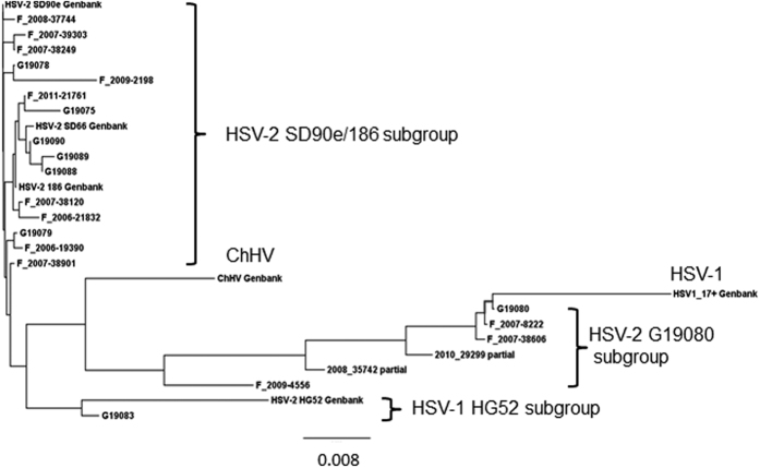
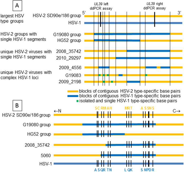
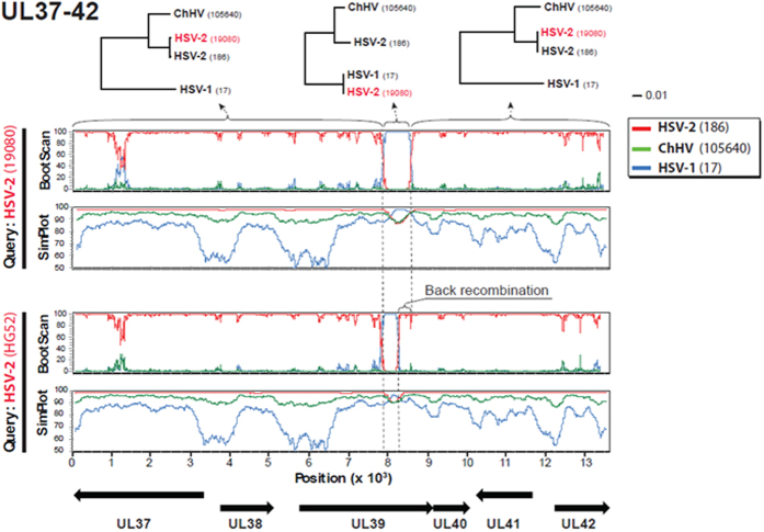
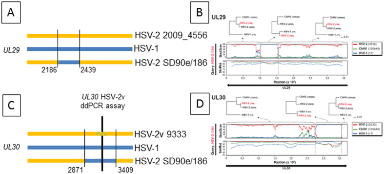
Homo sapiens harbor two distinct, medically significant species of simplexviruses, herpes simplex virus (HSV)-1 and HSV-2, with estimated divergence 6-8 million years ago (MYA). Unexpectedly, we found that circulating HSV-2 strains can contain HSV-1 DNA segments in three distinct genes. Using over 150 genital swabs from North and South America and Africa, we detected recombinants worldwide. Common, widely distributed gene UL39 genotypes are parsimoniously explained by an initial >457 basepair (bp) HSV-1 × HSV-2 crossover followed by back-recombination to HSV-2. Blocks of >244 and >539 bp of HSV-1 DNA within genes UL29 and UL30, respectively, have reached near fixation, with a minority of strains retaining sequences we posit as ancestral HSV-2. Our data add to previous in vitro and animal work, implying that in vivo cellular co-infection with HSV-1 and HSV-2 yields viable interspecies recombinants in the natural human host.


Conflict of interest statement

The authors declare no competing financial interests.

Figures

Figure 1
Figure 1. Phylogenetic tree of HSV-2 UL39 sequences from bp 950 to the stop codon.
The tree contains only one representative sequence for each unique nucleotide sequences in this region. Strain 333 is not included because it’s sequence is identical to SD90e in this region. Major subgroups, HSV-1 strain 17+, ChHV, and prototype strains from Genbank are identified. The largest HSV-2 group is contains the proposed prototype SD90e strain and strain 186, used for crossover analyses as described in the text.
Figure 2
Figure 2. HSV UL39 variants in circulating strains.
Horizontal lines represent C-terminal UL39 sequences to approximate scale. Group and virus names at left. (A) Genotypes. At top, the largest HSV-2 clade similar to strains SD90e and 186 (top) is yellow and the HSV-1 group is blue. Within IRV, blue bars represent zones of 4 or more contiguous HSV-1 variant SNPs. Green spots are isolated SNPs containing one HSV-1 variant nucleotide. Thin vertical black lines represent short crossover zones indistinguishable between HSV genotypes. Thick vertical black lines show approximate locations of type-specific ddPCR genotyping assays. (B) Amino acid variations in selected groups and viruses. Strains SD90e and 186 (top) are yellow and HSV-1 group (bottom) is blue. Blue bars in IRV represent zones of contiguous SNPS with HSV-1 variant nucleotides. Short black lines represent locations of color coded amino acid differences between strains.
Figure 3
Figure 3. Recombination analysis of the UL37 to UL42 genomic region in HSV-2 strains 19080 (upper) and HG52 (lower).
Bootscan and a Simplot analyses are depicted for each strain. At bottom the coding directions of the ORFs and length of the genomic region in kilobases from the stop codons in UL37 and UL42 are indicated. Clear shifts in bootstrap values supporting different phylogenetic topologies indicate recombination crossovers in the UL39 gene in both strains (also indicated by vertical dotted lines). These crossovers are supported by the Simplot analysis, which demonstrates a shift in similarity with a higher similarity to HSV-1 in the recombination fragment. To further test and visualize ancestry of the recombination fragment in strain 19080, phylogenetic trees based on the recombination fragment and flanking regions are shown. Strain 19080 clearly clusters closely to HSV-1 in the tree based on the recombination fragment, and closely to HSV-2 in the flanking regions, further supporting recombination with HSV-2 as major parental, and HSV-1 as minor parental strains. The shorter recombination fragment in HSV-2 strain HG52 suggests a back recombination, where a strain with the larger HSV-1 recombination fragment has recombined again with another HSV-2 strain and expelled a part of the recombinant fragment.
Figure 4
Figure 4. HSV UL29 and UL30 genotypes in strains circulating in humans.
(A and C) schematic diagrams of C-terminal coding sequences to approximate scale. HSV-1 is blue, rare HSV-2 strains are yellow. Blue bars in HSV-2 SD90e/186 represent circulating HSV-2 strains that have identity to HSV-1 in this region. Thin vertical black lines and associated strain 186 nucleotide numbers mark lateral flanks of HSV-1 identity. For UL30, thick vertical bar and yellow/green hatched zone within HSV-2v 9333 UL30 represent the locus detected by a ddPCR assay for which strain HSV-2v 9333 contains a variant nucleotide. (B,D) Recombination analysis of the UL29 and UL30 genes in HSV-2 strain 186. Bootscan and Simplot analyses are depicted for each gene. Clear shifts in bootstrap values supporting different phylogenetic topologies indicate recombination crossovers in both genes (also indicated by dotted lines). These crossovers are supported by the Simplot analysis, which demonstrates a shift in similarity with a higher similarity to HSV-1 in the recombination fragments. To further test and visualize ancestry of the recombination fragments, phylogenetic trees based on the recombination fragment and flanking regions are shown. HSV-2 strain 186 clearly shifts from clustering closely to HSV-2 in the trees based on the flanking regions, to clustering closely to HSV-1 in the trees based on the recombination fragments. These results suggest recombination with HSV-2 as major parental, and HSV-1 as minor parental strains.

Similar articles

Cited by

References

    1. Wertheim J. O., Smith M. D., Smith D. M., Scheffler K. & Kosakovsky Pond S. L. Evolutionary origins of human herpes simplex viruses 1 and 2. Molecular biology and evolution 31, 2356–2364, doi: 10.1093/molbev/msu185 (2014). - DOI - PMC - PubMed
    1. Bertke A. S., Patel A. & Krause P. R. Herpes simplex virus latency-associated transcript sequence downstream of the promoter influences type-specific reactivation and viral neurotropism. Journal of virology 81, 6605–6613, doi: 10.1128/JVI.02701-06 (2007). - DOI - PMC - PubMed
    1. Morse L. S., Buchman T. G., Roizman B. & Schaffer P. A. Anatomy of herpes simplex virus DNA. IX. Apparent exclusion of some parental DNA arrangements in the generation of intertypic (HSV-1 × HSV-2) recombinants. Journal of virology 24, 231–248 (1977). - PMC - PubMed
    1. Norberg P. et al.. A genome-wide comparative evolutionary analysis of herpes simplex virus type 1 and varicella zoster virus. PLoS One 6, e22527, doi: 10.1371/journal.pone.0022527 (2011). - DOI - PMC - PubMed
    1. Burrel S. et al.. Genetic Diversity within Alphaherpesviruses: Characterization of a Novel Variant of Herpes Simplex Virus 2. Journal of virology 89, 12273–12283, doi: 10.1128/JVI.01959-15 (2015). - DOI - PMC - PubMed

Publication types