

Hyaluronan synthesis by developing cortical neurons in vitro
- PMID: 28287145
- PMCID: PMC5347017
- DOI: 10.1038/srep44135
Hyaluronan synthesis by developing cortical neurons in vitro
Abstract
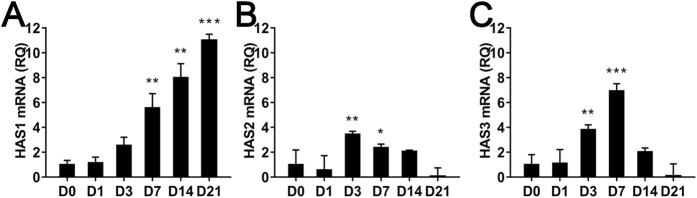
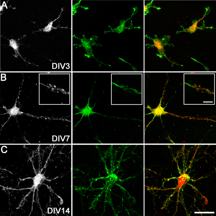
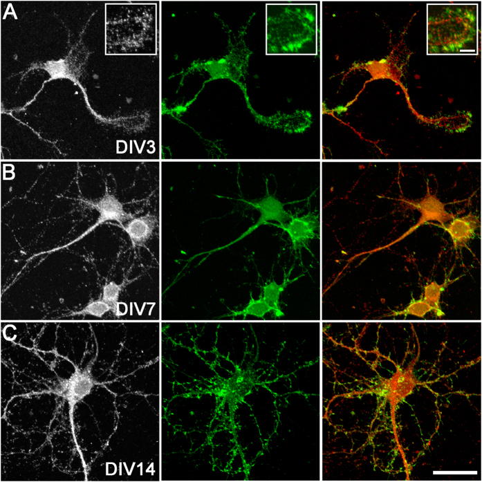
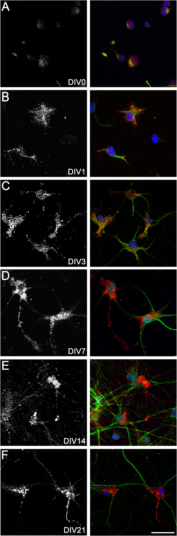
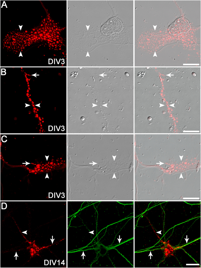
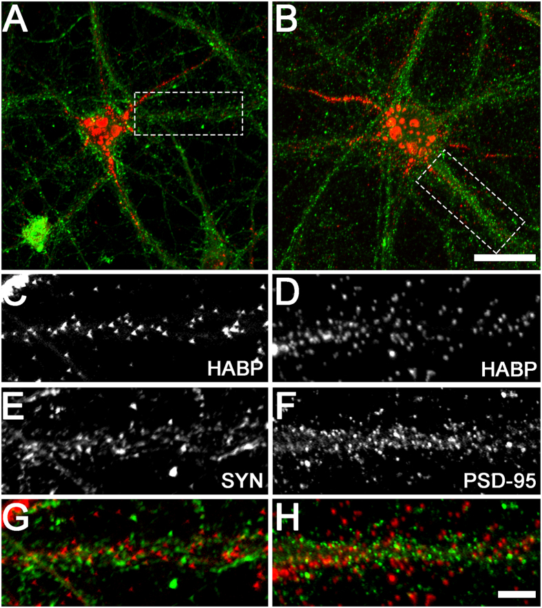
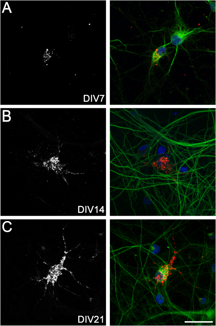
Hyaluronan is a linear glycosaminoglycan that forms the backbone of perineuronal nets around neurons in the cerebral cortex. However, it remains controversial whether neurons are capable of independent hyaluronan synthesis. Herein, we examined the expression of hyaluronan and hyaluronan synthases (HASs) throughout cortical neuron development in vitro. Enriched cultures of cortical neurons were established from E16 rats. Neurons were collected at days in vitro (DIV) 0 (4 h), 1, 3, 7, 14, and 21 for qPCR or immunocytochemistry. In the relative absence of glia, neurons exhibited HAS1-3 mRNA at all time-points. By immunocytochemistry, puncta of HAS2-3 protein and hyaluronan were located on neuronal cell bodies, neurites, and lamellipodia/growth cones from as early as 4 h in culture. As neurons matured, hyaluronan was also detected on dendrites, filopodia, and axons, and around synapses. Percentages of hyaluronan-positive neurons increased with culture time to ~93% by DIV21, while only half of neurons at DIV21 expressed the perineuronal net marker Wisteria floribunda agglutinin. These data clearly demonstrate that neurons in vitro can independently synthesise hyaluronan throughout all maturational stages, and that hyaluronan production is not limited to neurons expressing perineuronal nets. The specific structural localisation of hyaluronan suggests potential roles in neuronal development and function.
Conflict of interest statement
The authors declare no competing financial interests.
Figures
References
-
- Spicer A. P., Augustine M. L. & McDonald J. A. Molecular cloning and characterization of a putative mouse hyaluronan synthase. J. Biol. Chem. 271, 23400–23406 (1996). - PubMed
-
- Spicer A. P., Olson J. S. & McDonald J. A. Molecular cloning and characterization of a cDNA encoding the third putative mammalian hyaluronan synthase. J. Biol. Chem. 272, 8957–8961 (1997). - PubMed
-
- Watanabe K. & Yamaguchi Y. Molecular identification of a putative human hyaluronan synthase. J. Biol. Chem. 271, 22945–22948 (1996). - PubMed
-
- Laurent T. C., Laurent U. B. & Fraser J. R. The structure and function of hyaluronan: An overview. Immunol. Cell Biol. 74, A1–A7 (1996). - PubMed
-
- Toole B. P. Hyaluronan: from extracellular glue to pericellular cue. Nat. Rev. 4, 528–539 (2004). - PubMed
Publication types
MeSH terms
Substances
LinkOut - more resources
Full Text Sources
Other Literature Sources
