. 2017 Mar 13:7:44366.
doi: 10.1038/srep44366.

Crystal structures reveal N-terminal Domain of Arabidopsis thaliana ClpD to be highly divergent from that of ClpC1

Affiliations

Crystal structures reveal N-terminal Domain of Arabidopsis thaliana ClpD to be highly divergent from that of ClpC1

Chinmayee Mohapatra et al. Sci Rep. .

Abstract

The caseinolytic protease machinery associated chaperone protein ClpC is known to be present in bacteria, plants and other eukaryotes, whereas ClpD is unique to plants. Plant ClpC and ClpD proteins get localized into chloroplast stroma. Herein, we report high resolution crystal structures of the N-terminal domain of Arabidopsis thaliana ClpC1 and ClpD. Surprisingly, AtClpD, but not AtClpC1, deviates from the typical N-terminal repeat domain organization of known Clp chaperones and have only seven α-helices, instead of eight. In addition, the loop connecting the two halves of AtClpD NTD is longer and covers the region which in case of AtClpC1 is thought to contribute to adaptor protein interaction. Taken together, the N-terminal domain of AtClpD has a divergent structural organization compared to any known Clp chaperones which hints towards its specific role during plant stress conditions, as opposed to that in the maintenance of chloroplastic homeostasis by AtClpC1. Conservation of residues in the NTD that are responsible for the binding of the cyclic peptide activator - Cyclomarin A, as reported for mycobacterial ClpC1 suggests that the peptide could be used as an activator to both AtClpC1 and AtClpD, which could be useful in their detailed in vitro functional characterization.


Conflict of interest statement

The authors declare no competing financial interests.

Figures

Figure 1
Figure 1. Domain organization of AtClpC1 and AtClpD.
The organization of functional domains of AtClpC1 and AtClpD are shown with their respective amino acid residue boundaries. N corresponds to the N-terminal domain, D1 the first ATPase domain and D2 the second ATPase domain.
Figure 2
Figure 2. Structure of AtClpC1 N-terminal domain.
(A) Cartoon representation of the structure of A. thaliana ClpC1 NTD with the helices (blue) and loops (green). The N and C termini as well as the eight α-helices are labeled. (B) Structural alignment of AtClpC1 residues 99–161 (blue) with residues 174–236 (green).
Figure 3
Figure 3. Structure of AtClpD N-terminal domain.
(A) Cartoon representation of the structure of A. thaliana ClpD NTD with the helices (red) and loops (orange). The N and C termini as well as the seven α-helices are labeled. The four-residue long stretch in the long middle loop (that could not be seen in electron density) was fitted into the structure and is shown with intermittant gaps. (B) Structural alignment of AtClpD NTD (red helices and orange loops) with AtClpC1 NTD (blue helices and green loops). The N and C termini as well as α3 of AtClpC1 NTD are labeled.
Figure 4
Figure 4. Sequence alignment of AtClpD NTD with the NTD of putative ClpD from different plants.
The sequences have been aligned using ClustalW. For AtClpD, the residues forming helices are underlined and the helices are labelled α1 to α7. The residues conserved among all ten sequences are highlighted in red, those conserved among eight or nine sequences are highlighted in green and those conserved in five, six or seven sequences are highlighted in grey. The residues of regions that are substantially different in AtClpD structure as compared to ClpC have been given in blue. Bo, Cs, Gh, Nt, Rc, Vv, Os and Zm corresponds to Brassica oleracea, Citrus sinensis, Gossypium hirsutum, Nicotiana tabacum, Ricinus communis, Vitis vinifera, Oryza sativa and Zea mays respectively.
Figure 5
Figure 5. Electron density map for the loop region between helices α2 and α3 in the crystal structure of AtClpD.
The Fo-Fc omit map for the residues in the loop region between helices α2 and α3, contoured to 1.0 sigma is represented in grey colour. Helices and loop residues are labeled.
Figure 6
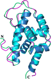
Figure 6. Structural alignment of AtClpC1 NTD with MtClpC1 NTD.
AtClpC1 NTD is shown in blue helices and green loops, MtClpC1 NTD is shown in cyan helices and magenta loops. The N and C termini are labeled.
Figure 7
Figure 7. Structure-based sequence alignment of AtClpC1 NTD and AtClpD NTD with bacterial ClpC NTDs.
The sequences have been aligned using ClustalW. The residues forming helices are underlined and the helices are labelled α1 to α8 (ClpC numbering). The residues conserved among all seven sequences are highlighted in red, those conserved among five or six sequences are highlighted in green and those conserved in four sequences are highlighted in grey. The residues of regions that are substantially different in AtClpD structure have been given in blue. Mt, Cg, Cd, Bs and Bl corresponds to Mycobacterium tuberculosis, Corynebacterium glutamicum, Clostridium difficile, Bacillus subtilis and Bacillus lehensis, respectively.
Figure 8
Figure 8. Structure-based sequence alignment of AtClpC1 NTD and AtClpD NTD with ClpA/B NTD and ClpT1/T2 proteins.
The sequences have been aligned using ClustalW and the residues forming helices are underlined and helices labelled α1 to α8 (ClpA/B/C/T numbering). The residues conserved among all seven sequences are highlighted in red, those conserved among five or six sequences are highlighted in green and those conserved among four sequences are highlighted in grey. The loop region of AtClpD corresponding to α3 of other structures is given in blue. Ec and Pf correspond to Escherichia coli and Plasmodium falciparum, respectively.
Figure 9
Figure 9. Structure-based sequence alignment of AtClpC1 NTD and AtClpD NTD with MtClpC1 NTD, highlighting the residues involved in Cyclomarin A interaction.
The sequences have been aligned using ClustalW and the residues forming helices are underlined. The residues of MtClpC1 NTD involved in Cyclomarin A interaction are shown in blue and with a star on top. The corresponding residues of AtClpC1 NTD and AtClpD NTD are shown in blue. The residues conserved among all three sequences are highlighted in red and those conserved among two sequences are highlighted in green.
Figure 10
Figure 10. Structural alignment of AtClpD NTD with MtClpC1 NTD in complex with Cyclomarin A.
MtClpC1 NTD is shown in cyan and residues involved in Cyclomarin A (CymA) interaction are shown in magenta (coordinates from PDB id: 3WDC). CymA is shown in stick model and in blue. AtClpD NTD is shown in red and the residues which could interact with CymA (Phe80, Phe168, Lys173 and Glu177) are shown as sticks in green and labelled. The region of NTDs coming in the proximity of CymA alone is shown for figure clarity.

Similar articles

Cited by

References

    1. Peltier J. B. et al.. Clp protease complexes from photosynthetic and non-photosynthetic plastids and mitochondria of plants, their predicted three-dimensional structures, and functional implications. J Biol Chem. 279, 4768–4781, doi: 10.1074/jbc.M309212200 (2004). - DOI - PubMed
    1. Peltier J. B., Ytterberg J., Liberles D. A., Roepstorff P. & van Wijk K. J. Identification of a 350-kDa ClpP protease complex with 10 different Clp isoforms in chloroplasts of Arabidopsis thaliana. J Biol Chem. 276, 16318–16327, doi: 10.1074/jbc.M010503200 (2001). - DOI - PubMed
    1. Adam Z. & Clarke A. K. Cutting edge of chloroplast proteolysis. Trends Plant Sci. 7, 451–456, doi: S1360-1385(02)02326-9 (2002). - PubMed
    1. Lee U. et al.. The Arabidopsis ClpB/Hsp100 family of proteins: chaperones for stress and chloroplast development. Plant J. 49, 115–127, doi: 10.1111/j.1365-313X.2006.02940.x (2007). - DOI - PubMed
    1. Mishra R. C. & Grover A. ClpB/Hsp100 proteins and heat stress tolerance in plants. Crit Rev Biotechnol. 1–13, doi: 10.3109/07388551.2015.1051942 (2015). - DOI - PubMed

Publication types

MeSH terms

Substances

LinkOut - more resources