

Enhanced osteogenesis and angiogenesis by mesoporous hydroxyapatite microspheres-derived simvastatin sustained release system for superior bone regeneration
- PMID: 28287178
- PMCID: PMC5347005
- DOI: 10.1038/srep44129
Enhanced osteogenesis and angiogenesis by mesoporous hydroxyapatite microspheres-derived simvastatin sustained release system for superior bone regeneration
Abstract
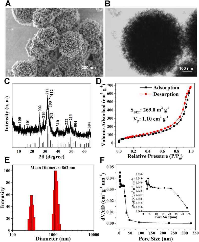
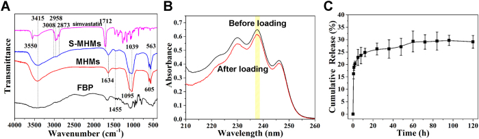
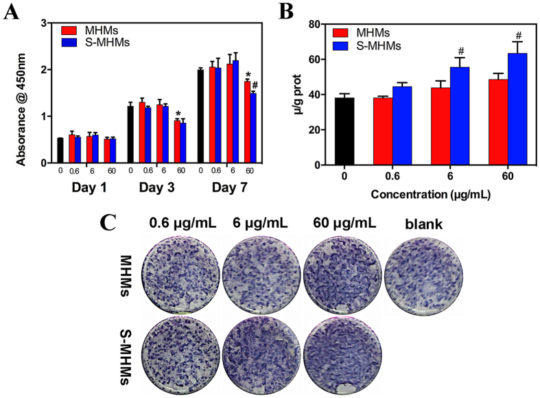
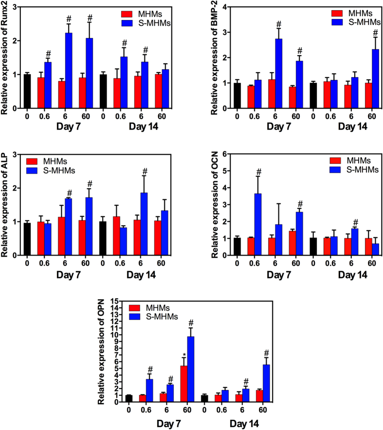
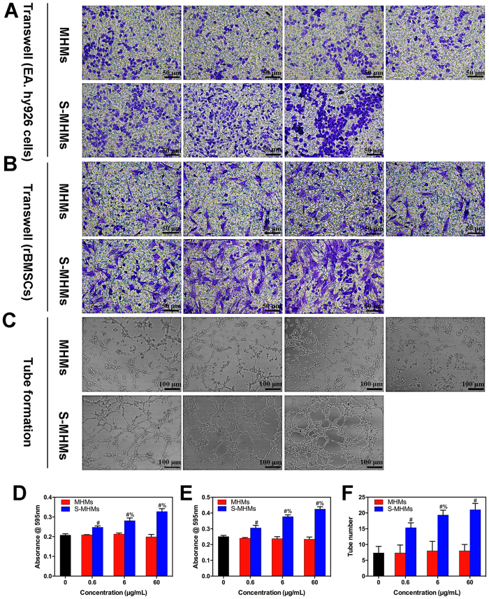
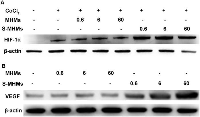
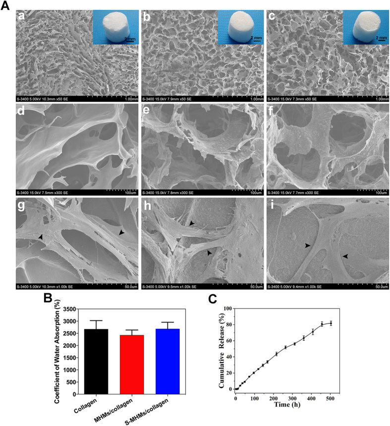
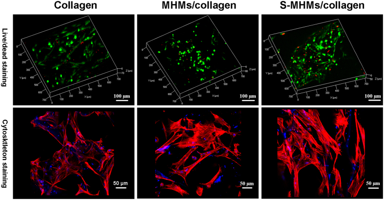
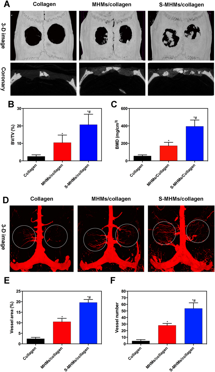
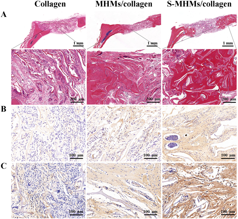
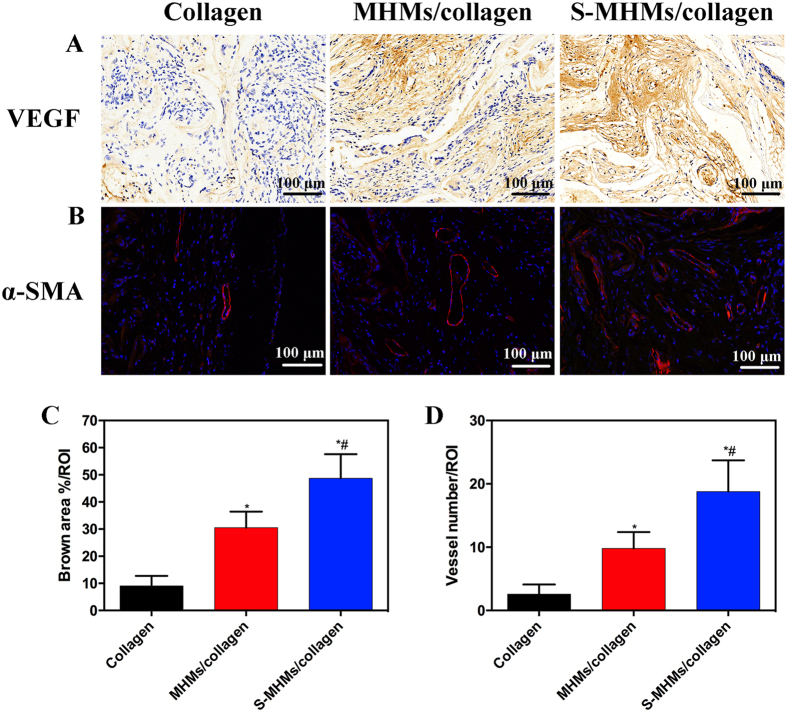
Biomaterials with both excellent osteogenic and angiogenic activities are desirable to repair massive bone defects. In this study, simvastatin with both osteogenic and angiogenic activities was incorporated into the mesoporous hydroxyapatite microspheres (MHMs) synthesized through a microwave-assisted hydrothermal method using fructose 1,6-bisphosphate trisodium salt (FBP) as an organic phosphorous source. The effects of the simvastatin-loaded MHMs (S-MHMs) on the osteogenic differentiation of rat bone marrow mesenchymal stem cells (rBMSCs) and angiogenesis in EA.hy926 cells were investigated. The results showed that the S-MHMs not only enhanced the expression of osteogenic markers in rBMSCs but also promoted the migration and tube formation of EA.hy926 cells. Furthermore, the S-MHMs were incorporated into collagen matrix to construct a novel S-MHMs/collagen composite scaffold. With the aid of MHMs, the water-insoluble simvastatin was homogenously incorporated into the hydrophilic collagen matrix and presented a sustained release profile. In vivo experiments showed that the S-MHMs/collagen scaffolds enhanced the bone regeneration and neovascularization simultaneously. These results demonstrated that the water-insoluble simvastatin could be incorporated into the MHMs and maintained its biological activities, more importantly, the S-MHMs/collagen scaffolds fabricated in this study are of immense potential in bone defect repair by enhancing osteogenesis and angiogenesis simultaneously.
Conflict of interest statement
The authors declare no competing financial interests.
Figures
Similar articles
-
Evaluation of zinc-doped mesoporous hydroxyapatite microspheres for the construction of a novel biomimetic scaffold optimized for bone augmentation.Int J Nanomedicine. 2017 Mar 24;12:2293-2306. doi: 10.2147/IJN.S126505. eCollection 2017. Int J Nanomedicine. 2017. PMID: 28392688 Free PMC article.
-
Copper-doped mesoporous hydroxyapatite microspheres synthesized by a microwave-hydrothermal method using creatine phosphate as an organic phosphorus source: application in drug delivery and enhanced bone regeneration.J Mater Chem B. 2017 Feb 7;5(5):1039-1052. doi: 10.1039/c6tb02747d. Epub 2017 Jan 18. J Mater Chem B. 2017. PMID: 32263882
-
Surface-decorated hydroxyapatite scaffold with on-demand delivery of dexamethasone and stromal cell derived factor-1 for enhanced osteogenesis.Mater Sci Eng C Mater Biol Appl. 2018 Aug 1;89:355-370. doi: 10.1016/j.msec.2018.04.008. Epub 2018 Apr 11. Mater Sci Eng C Mater Biol Appl. 2018. PMID: 29752108
-
Simvastatin-Incorporated Drug Delivery Systems for Bone Regeneration.ACS Biomater Sci Eng. 2021 Jun 14;7(6):2177-2191. doi: 10.1021/acsbiomaterials.1c00462. Epub 2021 Apr 20. ACS Biomater Sci Eng. 2021. PMID: 33877804 Review.
-
[Research progress on controlled release of various growth factors in bone regeneration].Zhongguo Xiu Fu Chong Jian Wai Ke Za Zhi. 2019 Jun 15;33(6):750-755. doi: 10.7507/1002-1892.201901116. Zhongguo Xiu Fu Chong Jian Wai Ke Za Zhi. 2019. PMID: 31198005 Free PMC article. Review. Chinese.
Cited by
-
Combined Delivery of Two Different Bioactive Factors Incorporated in Hydroxyapatite Microcarrier for Bone Regeneration.Tissue Eng Regen Med. 2020 Oct;17(5):607-624. doi: 10.1007/s13770-020-00257-5. Epub 2020 Aug 16. Tissue Eng Regen Med. 2020. PMID: 32803541 Free PMC article.
-
Potential bioactive coating system for high-performance absorbable magnesium bone implants.Bioact Mater. 2021 Oct 27;12:42-63. doi: 10.1016/j.bioactmat.2021.10.034. eCollection 2022 Jun. Bioact Mater. 2021. PMID: 35087962 Free PMC article. Review.
-
Silver Nanoparticles and Simvastatin-Loaded PLGA-Coated Hydroxyapatite/Calcium Carbonate Scaffolds.Nanomaterials (Basel). 2024 Oct 12;14(20):1637. doi: 10.3390/nano14201637. Nanomaterials (Basel). 2024. PMID: 39452973 Free PMC article.
-
3D printed poly(ε-caprolactone) scaffolds function with simvastatin-loaded poly(lactic-co-glycolic acid) microspheres to repair load-bearing segmental bone defects.Exp Ther Med. 2019 Jan;17(1):79-90. doi: 10.3892/etm.2018.6947. Epub 2018 Nov 9. Exp Ther Med. 2019. PMID: 30651767 Free PMC article.
-
Physical and drug- releasing properties of a cement containing simvastatin (SimCeram).BMC Oral Health. 2025 May 5;25(1):684. doi: 10.1186/s12903-025-06045-8. BMC Oral Health. 2025. PMID: 40325441 Free PMC article.
References
-
- Dickson K. F., Katzman S. & Paiement G. The importance of the blood supply in the healing of tibial fractures. Contemp. Orthop. 30, 489–493 (1995). - PubMed
-
- Stegen S., van Gastel N. & Carmeliet G. Bringing new life to damaged bone: the importance of angiogenesis in bone repair and regeneration. Bone 70, 19–27 (2015). - PubMed
-
- Sonobe M. et al.. Stimulatory effects of statins on bone marrow-derived mesenchymal stem cells. Study of a new therapeutic agent for fracture. Bio-med. Mater. Eng. 15, 261–267 (2005). - PubMed
-
- Song C. et al.. Simvastatin induces osteoblastic differentiation and inhibits adipocytic differentiation in mouse bone marrow stromal cells. Biochem. Biophys. Res. Commun. 308, 458–462 (2003). - PubMed
-
- Ruiz-Gaspa S. et al.. Simvastatin and atorvastatin enhance gene expression of collagen type 1 and osteocalcin in primary human osteoblasts and MG-63 cultures. J. Cell. Biochem. 101, 1430–1438 (2007). - PubMed
Publication types
MeSH terms
Substances
LinkOut - more resources
Full Text Sources
Other Literature Sources
Miscellaneous
