

doi: 10.1051/sicotj/2017010.
Epub 2017 Mar 10.
Management of soft-tissue sarcomas; treatment strategies, staging, and outcomes
Affiliations
- PMID: 28287387
- PMCID: PMC5347369
- DOI: 10.1051/sicotj/2017010
Item in Clipboard
Management of soft-tissue sarcomas; treatment strategies, staging, and outcomes
SICOT J.
2017.
Abstract
Soft-tissue sarcomas (STS) are a rare group of malignant tumors which can affect any age group. For the majority of patients who present with a localized STS, treatment involves a multidisciplinary team decision-making approach ultimately relying on surgical resection with or without adjuvant radiation for successful limb salvage. The goals of treatment are to provide the patient with a functional extremity without local tumor relapse. The purpose of this article is to review the treatment of extremity STS, with a focus on staging, treatment options, and outcomes.
© The Authors, published by EDP Sciences, 2017.
Figures
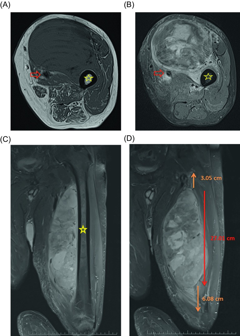
Selected T1 (A) and fat-saturated T2 (B) axial as well as fat-saturated coronal T2 (C) MRI images of a 60-year-old patient with a large, deep mass located in the anterior thigh. On the pretreatment imaging the mass was intimately associated with the femoral neurovascular bundle (arrow) as well as the periosteum of the femur (star). A biopsy was performed and showed high-grade pleomorphic rhabdomyosarcoma. The mass measured approximately 27 cm cranial/caudal however was associated with peritumoral edema which spanned nearly the entire length of the femur on coronal fat-saturated T2 (D) MRI images.
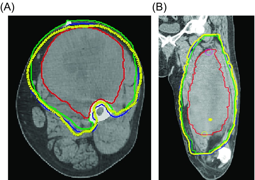
Preoperative radiotherapy planning volumes for the patient in Figure 1 are shown on axial (A) and coronal (B) CT images. The Gross Tumor Volume (GTV) is demonstrated by the solid red contour; Clinical Target Volume (CTV) is demonstrated by the green solid contour; Planning Target Volume (PTV) is shown by the blue solid contour; and the thick yellow line represents the prescribed radiotherapy dose volume. Note that intensity-modulated radiotherapy (IMRT) was used to adequately encompass the radiotherapy target volume while avoiding the bone by sculpting the high dose volume around the femoral cortex for protection purposes (A), while also accounting for the peritumoral edema surrounding the lesion (B) which was demonstrated on the coronal fat-saturated T2 post-gadolinium image in Figures 1C and 1D.
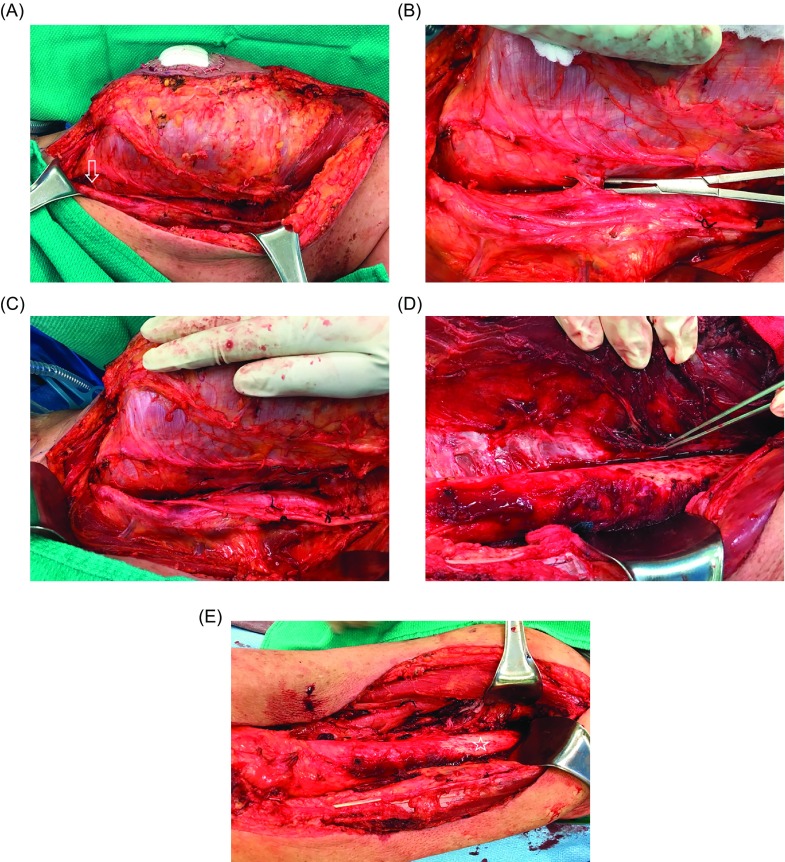
At the time of surgical excision (A), the femoral neurovascular bundle was very close to the tumor (arrow), with multiple perforating blood vessels entering the tumor (B). Due to preoperative IMRT it was safe to create a dissection plane between the tumor and the neurovascular bundle (C). The periosteum was also raised from the femur (pointer) as a margin along the tumor in the region where it was adherent to the bone (D). Although preoperative imaging showed the tumor to be very close to bone along the entire length of the femur, it was actually adherent to bone over a shorter length, so that only a small portion of the periosteum had to be removed (star) from the femoral shaft (E). The final pathological tumor resection margins were negative.
References
-
- Siegel RL, Miller KD, Jemal A (2015) Cancer statistics, 2015. CA Cancer J Clin 65(1), 5–29. - PubMed
-
- Pollock RE, Karnell LH, Menck HR, Winchester DP (1996) The National Cancer Data Base report on soft tissue sarcoma. Cancer 78(10), 2247–2257. - PubMed
-
- Fletcher CD (2014) The evolving classification of soft tissue tumours – an update based on the new 2013 WHO classification. Histopathology 64(1), 2–11. - PubMed
-
- Gerrand CH, Bell RS, Wunder JS, Kandel RA, O’Sullivan B, Catton CN, Griffin AM, Davis AM (2003) The influence of anatomic location on outcome in patients with soft tissue sarcoma of the extremity. Cancer 97(2), 485–492. - PubMed
-
- Frassica FJ, Khanna JA, McCarthy EF (2000) The role of MR imaging in soft tissue tumor evaluation: perspective of the orthopedic oncologist and musculoskeletal pathologist. Magn Reson Imaging Clin N Am 8(4), 915–927. - PubMed
LinkOut - more resources
Full Text Sources
Other Literature Sources