Review
. 2017 Mar 10;18(3):606.
doi: 10.3390/ijms18030606.

Keloid and Hypertrophic Scars Are the Result of Chronic Inflammation in the Reticular Dermis

Affiliations
Review

Keloid and Hypertrophic Scars Are the Result of Chronic Inflammation in the Reticular Dermis

Rei Ogawa. Int J Mol Sci. .

Abstract

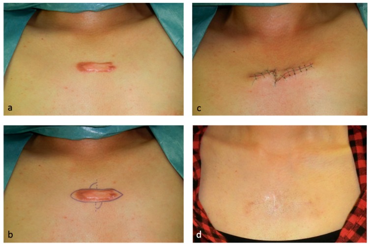
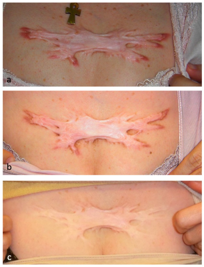
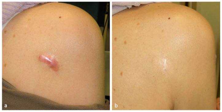
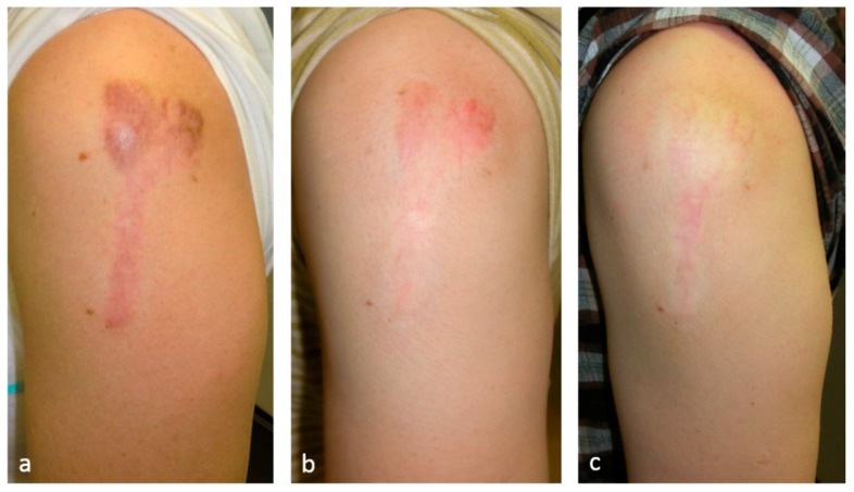
Keloids and hypertrophic scars are caused by cutaneous injury and irritation, including trauma, insect bite, burn, surgery, vaccination, skin piercing, acne, folliculitis, chicken pox, and herpes zoster infection. Notably, superficial injuries that do not reach the reticular dermis never cause keloidal and hypertrophic scarring. This suggests that these pathological scars are due to injury to this skin layer and the subsequent aberrant wound healing therein. The latter is characterized by continuous and histologically localized inflammation. As a result, the reticular layer of keloids and hypertrophic scars contains inflammatory cells, increased numbers of fibroblasts, newly formed blood vessels, and collagen deposits. Moreover, proinflammatory factors, such as interleukin (IL)-1α, IL-1β, IL-6, and tumor necrosis factor-α are upregulated in keloid tissues, which suggests that, in patients with keloids, proinflammatory genes in the skin are sensitive to trauma. This may promote chronic inflammation, which in turn may cause the invasive growth of keloids. In addition, the upregulation of proinflammatory factors in pathological scars suggests that, rather than being skin tumors, keloids and hypertrophic scars are inflammatory disorders of skin, specifically inflammatory disorders of the reticular dermis. Various external and internal post-wounding stimuli may promote reticular inflammation. The nature of these stimuli most likely shapes the characteristics, quantity, and course of keloids and hypertrophic scars. Specifically, it is likely that the intensity, frequency, and duration of these stimuli determine how quickly the scars appear, the direction and speed of growth, and the intensity of symptoms. These proinflammatory stimuli include a variety of local, systemic, and genetic factors. These observations together suggest that the clinical differences between keloids and hypertrophic scars merely reflect differences in the intensity, frequency, and duration of the inflammation of the reticular dermis. At present, physicians cannot (or at least find it very difficult to) control systemic and genetic risk factors of keloids and hypertrophic scars. However, they can use a number of treatment modalities that all, interestingly, act by reducing inflammation. They include corticosteroid injection/tape/ointment, radiotherapy, cryotherapy, compression therapy, stabilization therapy, 5-fluorouracil (5-FU) therapy, and surgical methods that reduce skin tension.

Keywords: hypertrophic scar; keloid; radiation; steroid tape; surgery.


Conflict of interest statement

The author declares no conflict of interest.

Figures

Figure 1
Figure 1
This 24-year-old female had a minor acne keloid on her chest that was treated by surgery, namely, total excision and z-plasty, plus postoperative taping fixation. Since the keloid was small, it could be removed totally and the tension was released by the z-plasty. Immediately after removing the sutures on day 10, silicone tape fixation was started to stabilize the wound. Eighteen months after surgery, recurrence was not observed. (a) Preoperative view; (b) design of the z-plasty; (c) immediately after surgery; and (d) 18 months after the operation.
Figure 2
Figure 2
This 51-year-old female had a minor chest wall keloid that was treated by high-dose-rate superficial brachytherapy. A total of 25 Gy was administered in five fractions over 5 days. The inflammation resolved completely. After 1 year of treatment, both the subjective and objective symptoms had improved dramatically. (a) Pretreatment view; (b) 6 months post-treatment; and (c) 4 years post-treatment.
Figure 3
Figure 3
This 75-year-old female had a mild right scapular keloid that was treated by deprodone propionate tape (Eclar® plaster). The tape was placed on the keloid 24 h a day and was changed daily. The inflammation resolved completely. After 19 months of treatment, both the subjective and objective symptoms of the patient had improved dramatically. (a) Pretreatment view; and (b) after 19 months of treatment.
Figure 4
Figure 4
This 60-year-old female had a mild keloid that was treated by 1064 nm long-pulsed Nd:YAG laser. Long-pulsed 1064 nm Nd:YAG laser was used at the following settings: 5 mm spot diameter, 75 J/cm2, 25 msec, and 2 Hz. After 1 year of treatment, the scar had almost disappeared. The known target of long-pulsed Nd:YAG lasers is the blood vessels. Thus, we speculate that laser therapy largely treats pathological scars by decreasing blood vessel numbers, thereby inhibiting the inflammatory signals coming from the blood vessels. (a) Pretreatment view; (b) after 10 months of treatment; and (c) after 16 months of treatment.

Similar articles

Cited by

References

    1. Huang C., Akaishi S., Hyakusoku H., Ogawa R. Are keloid and hypertrophic scar different forms of the same disorder? A fibroproliferative skin disorder hypothesis based on keloid findings. Int. Wound J. 2014;11:517–522. doi: 10.1111/j.1742-481X.2012.01118.x. - DOI - PMC - PubMed
    1. Ogawa R., Akaishi S. Endothelial dysfunction may play a key role in keloid and hypertrophic scar pathogenesis—Keloids and hypertrophic scars may be vascular disorders. Med. Hypotheses. 2016;96:51–60. doi: 10.1016/j.mehy.2016.09.024. - DOI - PubMed
    1. Deitch E.A., Wheelahan T.M., Rose M.P., Clothier J., Cotter J. Hypertrophic burn scars: Analysis of variables. J. Trauma. 1983;23:895–898. doi: 10.1097/00005373-198310000-00009. - DOI - PubMed
    1. Huang C., Liu L., You Z., Wang B., Du Y., Ogawa R. Keloid progression: A stiffness gap hypothesis. Int. Wound J. 2016 doi: 10.1111/iwj.12693. - DOI - PMC - PubMed
    1. Akaishi S., Akimoto M., Ogawa R., Hyakusoku H. The relationship between keloid growth pattern and stretching tension: Visual analysis using the finite element method. Ann. Plast. Surg. 2008;60:445–451. doi: 10.1097/SAP.0b013e3181238dd7. - DOI - PubMed