. 2017 Mar 13;6(3):e301.
doi: 10.1038/oncsis.2017.8.

Overexpression of SMC4 activates TGFβ/Smad signaling and promotes aggressive phenotype in glioma cells

Affiliations

Overexpression of SMC4 activates TGFβ/Smad signaling and promotes aggressive phenotype in glioma cells

L Jiang et al. Oncogenesis. .

Erratum in

Abstract

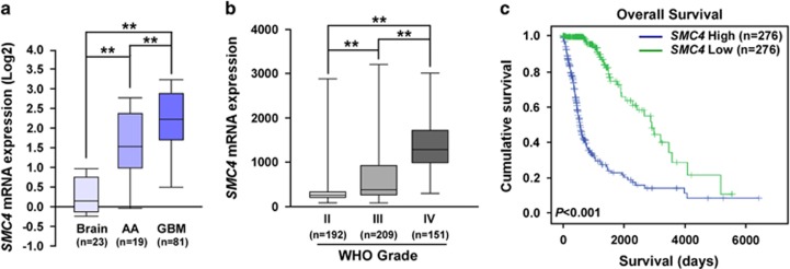
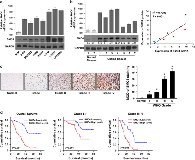
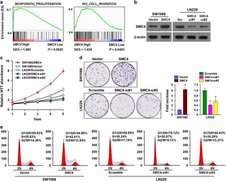
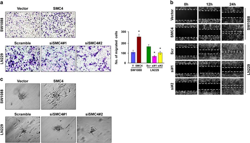
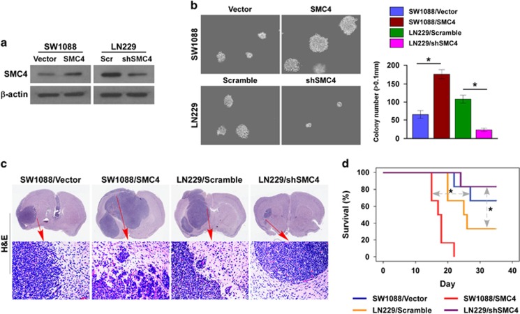
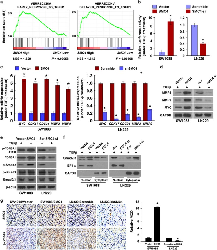
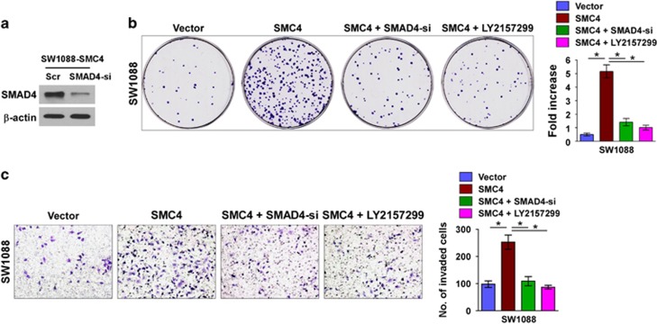
Overexpression of structural maintenance of chromosomes 4 (SMC4) has been reported to be involved in tumor cell growth, migration and invasion, and to be correlated with poor prognosis of cancer patient. However, its clinical significance and biological role in glioma remain unknown. Herein, we found that SMC4 expression at both mRNA and protein level was markedly increased in glioma cells and clinical tissues and that it correlated with poor prognosis. SMC4 overexpression markedly promoted the glioma cell proliferation rate and migration and invasive capability in vitro and in vivo, whereas SMC4 downregulation reduced it. Moreover, the transforming growth factor β (TGFβ)/Smad signaling pathway, which was activated in SMC4-transduced glioma cells and inhibited in SMC4-silenced glioma cells, contributed to SMC4-mediated glioma cell aggressiveness. Our results provide new insight into the oncofunction of SMC4 and the mechanism by which the TGFβ/Smad pathway is hyperactivated in gliomas, indicating that SMC4 is a valuable prognostic factor and a potential therapeutic target in gliomas.


Conflict of interest statement

The authors declare no conflict of interest.

Figures

Figure 1
Figure 1
High SMC4 mRNA expression in human glioma correlates with poor prognosis. (a) Quantification analysis of SMC4 mRNA expression in normal brain tissues, anaplastic astrocytoma (AA), and GBM specimens from the Oncomine database (GSE4290 specimens). (b) Quantification analysis of SMC4 mRNA expression in 522 glioma specimens of different WHO grades from TCGA. (c) Correlation analysis of SMC4 mRNA expression and overall survival of 522 patients with glioma from TCGA. **P<0.01.
Figure 2
Figure 2
SMC4 protein overexpression correlates with poor prognosis in human glioma. (a) Real-time reverse transcription–PCR (RT-PCR) and western blot detection of SMC4 mRNA and protein expression in NHA and cultured glioma cell lines (A172, SW1088, T98G, Hs683, LN18, LN229 and U118MG). (b) Real-time RT-PCR and western blot detection of SMC4 mRNA and protein expression in two normal brain tissue samples and seven glioma tissue samples (left). The Spearman correlation coefficient was calculated to assess the significance of association between SMC4 mRNA and protein expression levels (right, P<0.001). (c) IHC analysis of SMC4 protein expression in 194 glioma specimens of different WHO grades (left), magnification, × 400. Statistical quantification of the average mean optical densities (MODs) of SMC4 staining of 194 glioma specimens of different WHO grades (right). (d) Kaplan–Meier analysis of SMC4 expression levels in WHO grades I–IV gliomas and patient survival. *P<0.05.
Figure 3
Figure 3
Elevated SMC4 expression promotes glioma cell proliferation and viability in vitro. (a) GSEA plot indicating a significant correlation between SMC4 mRNA expression level and the proliferation and migration gene signatures (BENPORATH_PROLIFERATION, WU_CELL_MIGRATION). (b) Western blot detection of SMC4 protein expression in SW1088 and LN229 cells. (c) MTT assay of SW1088 and LN229 cell growth curves following SMC4 or SMC4 siRNA(s) transfection. (d) Representative micrographs (left) and quantification (right) of crystal violet–stained SW1088 and LN229 cell colonies following 14-day colony formation assay. (e) Flow cytometric analysis of cellular DNA synthesis and cell cycle progression in SW1088 and LN229 cells. Bars represent the mean±s.d. of three independent experiments. *P<0.05. Scr, scramble.
Figure 4
Figure 4
SMC4 promotes glioma cell migration and invasive capability in vitro. (a) Representative images (left, magnification, × 200) and quantification (right) of SW1088 and LN229 cell migration in the Transwell assay. The quantification of migrated cells is the mean of three independent experiments. Bars represent the mean±s.d. of three independent experiments. *P<0.05. (b) Wound-healing assay assessment of cell migration. (c) Representative micrographs of SW1088 and LN229 cells after 10-day culture in three-dimensional spheroid invasion assays, magnification, × 200. Scr, scramble.
Figure 5
Figure 5
SMC4 accelerates glioma cell tumorigenicity in vivo. (a) Western blot validation of SW1088 cells stably expressing SMC4 and LN229 cells stably expressing SMC4 shRNA. (b) Representative micrographs (left) and quantification (right) of colonies >0.1 mm formed in the anchorage-independent growth assay. (c) Intracranial brain tumor xenograft model in nude mice; representative images of tumors from each group are shown. Hematoxylin–eosin (H&E, lower panel magnification, × 100) staining demonstrated that SMC4 overexpression induced the aggressive phenotype of glioma cells in vivo, whereas SMC4 suppression inhibited it. (d) Kaplan–Meier survival analysis of the mice (n=6 per group). Bars represent the mean±s.d. of three independent experiments. *P<0.05.
Figure 6
Figure 6
SMC4 promotes glioma cell aggressiveness by activating the TGFβ/Smad signaling pathway. (a) GSEA plot indicating a significant correlation between SMC4 mRNA expression levels and early TGFβ-activated and delayed TGF-induced TGFβ gene signatures. (b) Luciferase-reported Smad activity in SW1088 and LN229 cells that were serum starved for 12 h before treatment with TGFβ (100 pM). (c) RT-PCR detection of MYC, CDK17, CDC34, MMP2 and MMP9 gene expression in SW1088 and LN229 cells that were serum starved for 12 h before treatment with TGFβ (100 pM). (d) Western blot detection of MMP2, MMP9 and MYC protein expression in SW1088 and LN229 cells that were serum starved for 12 h before treatment with TGFβ (100 pM). (e) Western blot detection of p-TGFBR1, TGFBR1, p-Smad2, p-Smad3 and Smad2/3 in the indicated cells that were serum starved for 12 h before treatment with TGFβ (100 pM). (f) Western blot detection of Smad2/3 expression levels in the nucleus or cytoplasm of SW1088 and LN229 cells treated with TGFβ (100 pM). (g) IHC staining (left) and quantification (right) of SMC4 and p-SMAD3 in the indicated tumor tissues. Magnification, × 400. Bars represent the mean±s.d. of three independent experiments. *P<0.05.
Figure 7
Figure 7
The TGFβ/Smad pathway contributes to SMC4-mediated aggressiveness of glioma cells. (a) Western blotting analysis of SMAD4 protein expression in SW1088 cells. (b) Representative micrographs (left) and quantification (right) of crystal violet-stained SW1088 cell colonies following 14-day colony formation assay. (c) Representative images (left, magnification, × 200) and quantification (right) of SW1088 cell invasion in the Transwell matrix penetration assay. Bars represent the mean±s.d. of three independent experiments. *P<0.05.

Similar articles

Cited by

References

    1. Ostrom QT, Gittleman H, Fulop J, Liu M, Blanda R, Kromer C et al. CBTRUS statistical report: primary brain and central nervous system tumors diagnosed in the United States in 2008-2012. Neuro-Oncology 2015; 17 (Suppl 4): iv1–iv62. - PMC - PubMed
    1. Louis DN, Perry A, Reifenberger G, von Deimling A, Figarella-Branger D, Cavenee WK et al. The 2016 World Health Organization classification of tumors of the central nervous system: a summary. Acta Neuropathol 2016; 131: 803–820. - PubMed
    1. Waghmare I, Roebke A, Minata M, Kango-Singh M, Nakano I. Intercellular cooperation and competition in brain cancers: lessons from Drosophila and human studies. Stem Cells Transl Med 2014; 3: 1262–1268. - PMC - PubMed
    1. Roesler R, Brunetto AT, Abujamra AL, de Farias CB, Brunetto AL, Schwartsmann G. Current and emerging molecular targets in glioma. Exp Rev Anticancer Ther 2010; 10: 1735–1751. - PubMed
    1. Gabayan AJ, Green SB, Sanan A, Jenrette J, Schultz C, Papagikos M et al. GliaSite brachytherapy for treatment of recurrent malignant gliomas: a retrospective multi-institutional analysis. Neurosurgery 2006; 58: 701–709; discussion 701–709. - PubMed