

The Effect of Contrast Material on Radiation Dose at CT: Part II. A Systematic Evaluation across 58 Patient Models
- PMID: 28287916
- PMCID: PMC5452877
- DOI: 10.1148/radiol.2017152852
The Effect of Contrast Material on Radiation Dose at CT: Part II. A Systematic Evaluation across 58 Patient Models
Abstract
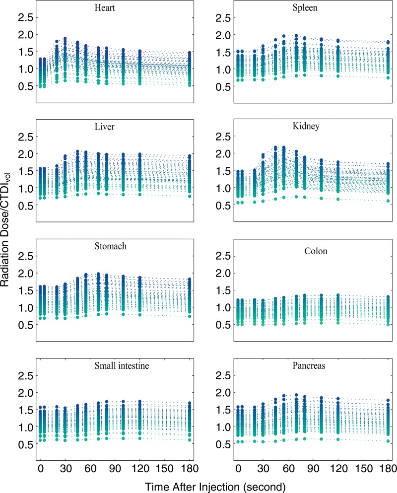
Purpose To estimate the radiation dose as a result of contrast medium administration in a typical abdominal computed tomographic (CT) examination across a library of contrast material-enhanced computational patient models. Materials and Methods In part II of this study, first, the technique described in part I of this study was applied to enhance the extended cardiac-torso models with patient-specific iodine-time profiles reflecting the administration of contrast material. Second, the patient models were deployed to assess the patient-specific organ dose as a function of time in a typical abdominal CT examination using Monte Carlo simulation. In this hypothesis-generating study, organ dose refers to the total energy deposited in the unit mass of the tissue inclusive of iodine. Third, a study was performed as a strategy to anticipate the biologically relevant dose (absorbed dose to tissue) in highly perfused organs such as the liver and kidney. The time-varying organ-dose increment values relative to those for unenhanced CT examinations were reported. Results The results from the patient models subjected to the injection protocol indicated up to a total 53%, 30%, 35%, 54%, 27%, 18%, 17%, and 24% increase in radiation dose delivered to the heart, spleen, liver, kidneys, stomach, colon, small intestine, and pancreas, respectively. The biologically relevant dose increase with respect to the dose at an unenhanced CT examination was in the range of 0%-18% increase for the liver and 27% for the kidney across 58 patient models. Conclusion The administration of contrast medium increases the total radiation dose. However, radiation dose, while relevant to be included in estimating the risk associated with contrast-enhanced CT, may still not fully characterize the total biologic effects. Therefore, given the fact that many CT diagnostic decisions would be impossible without the use of iodine, this study suggests the need to consider the effect of iodinated contrast material on the organ doses to patients undergoing CT studies when designing CT protocols. © RSNA, 2017 Online supplemental material is available for this article.
Figures


Comment in
-
The Effect of Iodine-based Contrast Material on Radiation Dose at CT: It's Complicated.Radiology. 2017 Jun;283(3):624-627. doi: 10.1148/radiol.2017170611. Radiology. 2017. PMID: 28514218 Free PMC article.
-
Effect of Iodine-based Contrast Material on Radiation Dose at CT.Radiology. 2017 Dec;285(3):1053-1054. doi: 10.1148/radiol.2017171523. Radiology. 2017. PMID: 29155625 Free PMC article. No abstract available.
References
-
- Cassola VF, Lima VJ, Kramer R, Khoury HJ. FASH and MASH: female and male adult human phantoms based on polygon mesh surfaces. I. Development of the anatomy. Phys Med Biol 2010;55(1):133–162. - PubMed
-
- Amato E, Salamone I, Naso S, Bottari A, Gaeta M, Blandino A. Can contrast media increase organ doses in CT examinations? a clinical study. AJR Am J Roentgenol 2013;200(6):1288–1293. - PubMed
-
- Garnica-Garza HM. Contrast-enhanced radiotherapy: feasibility and characteristics of the physical absorbed dose distribution for deep-seated tumors. Phys Med Biol 2009;54(18):5411–5425. - PubMed
Publication types
MeSH terms
Substances
Grants and funding
LinkOut - more resources
Full Text Sources
Other Literature Sources
Medical
