. 2017 May;13(5):514-521.
doi: 10.1038/nchembio.2329. Epub 2017 Mar 13.

Structural basis of PROTAC cooperative recognition for selective protein degradation

Affiliations

Structural basis of PROTAC cooperative recognition for selective protein degradation

Morgan S Gadd et al. Nat Chem Biol. 2017 May.

Abstract

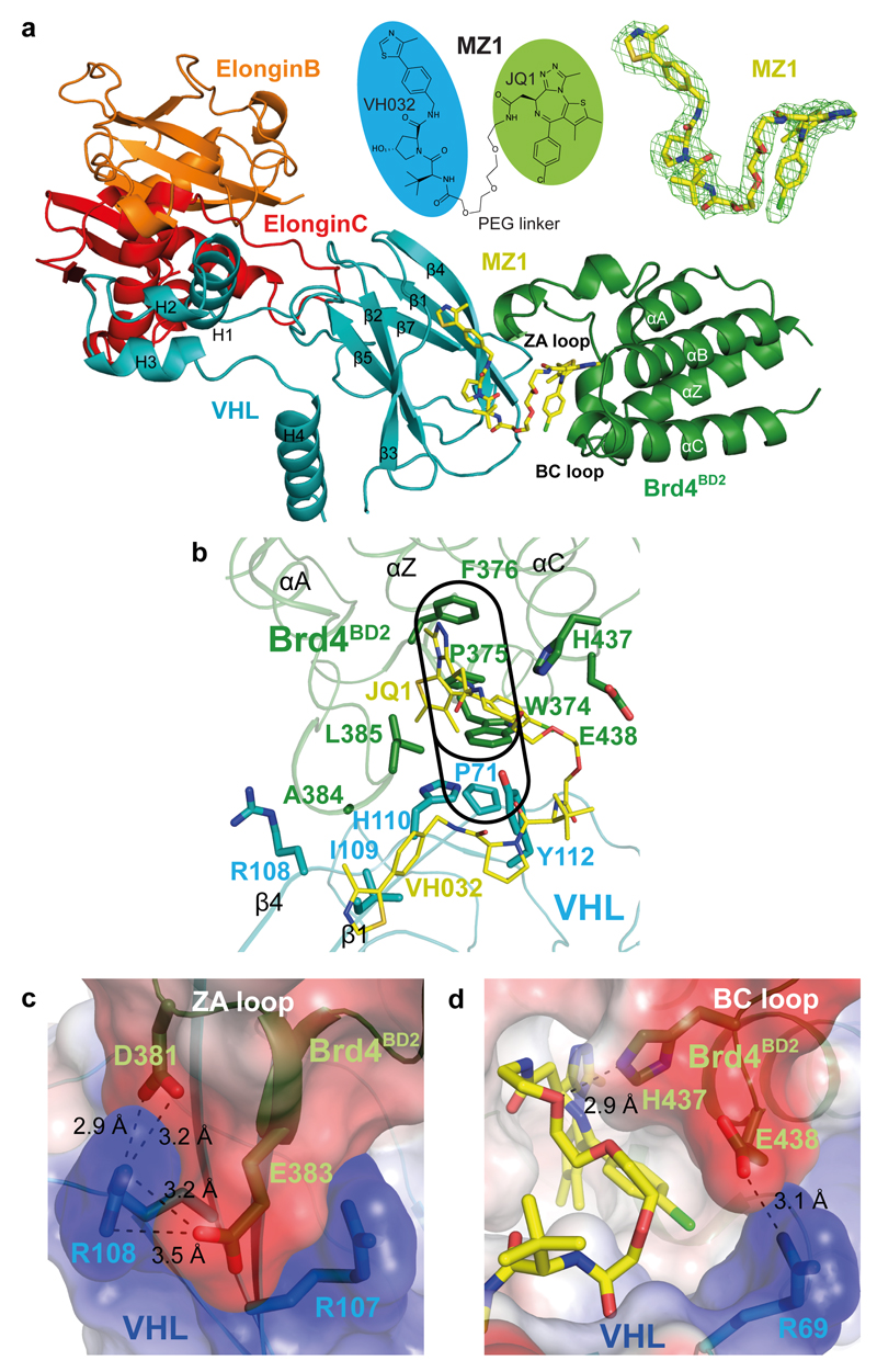
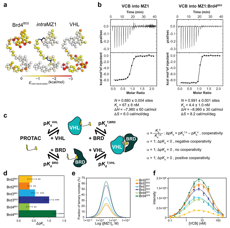
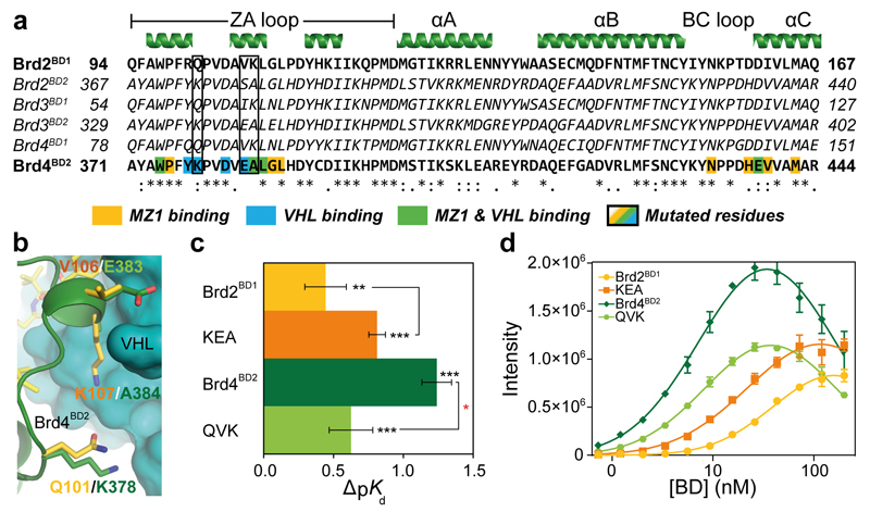
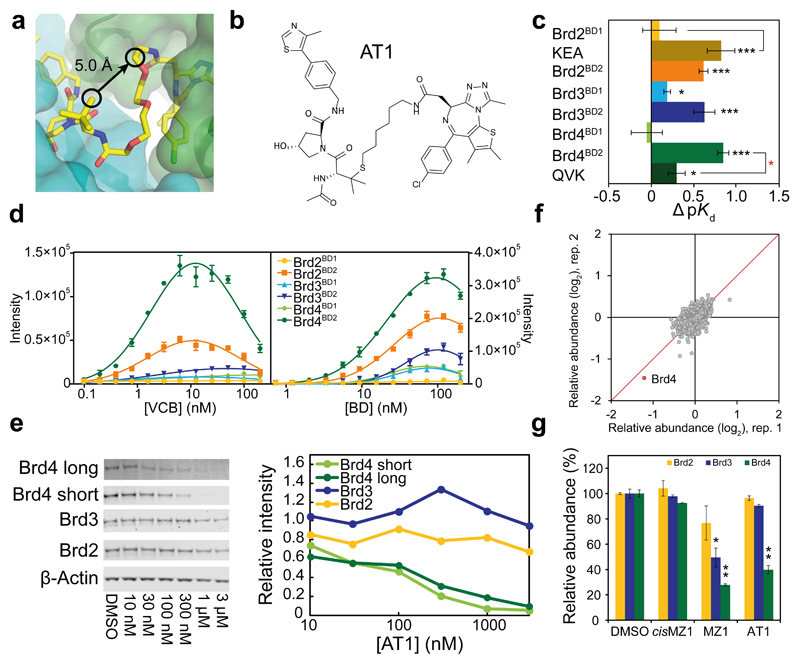
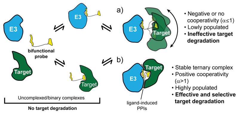
Inducing macromolecular interactions with small molecules to activate cellular signaling is a challenging goal. PROTACs (proteolysis-targeting chimeras) are bifunctional molecules that recruit a target protein in proximity to an E3 ubiquitin ligase to trigger protein degradation. Structural elucidation of the key ternary ligase-PROTAC-target species and its impact on target degradation selectivity remain elusive. We solved the crystal structure of Brd4 degrader MZ1 in complex with human VHL and the Brd4 bromodomain (Brd4BD2). The ligand folds into itself to allow formation of specific intermolecular interactions in the ternary complex. Isothermal titration calorimetry studies, supported by surface mutagenesis and proximity assays, are consistent with pronounced cooperative formation of ternary complexes with Brd4BD2. Structure-based-designed compound AT1 exhibits highly selective depletion of Brd4 in cells. Our results elucidate how PROTAC-induced de novo contacts dictate preferential recruitment of a target protein into a stable and cooperative complex with an E3 ligase for selective degradation.


Conflict of interest statement

Competing financial interests

The authors declare no competing financial interests.

Figures

Figure 1
Figure 1. The crystal structure of the Brd4BD2:MZ1:VHL-ElonginC-ElonginB complex.
a, Overall structure of Brd4BD2:MZ1:VHL-ElonginC-ElonginB in ribbon representation. Top middle, chemical structure of bifunctional PROTAC molecule MZ1. Top right, FoFc omit map generated prior to ligand modelling contoured at 3.0σ around bound MZ1. b, Key residues forming the hydrophobic “base” of the induced Brd4BD2:VHL interface. The "WPF" shelf of Brd4BD2 and extended "PWPF" stack are outlined in black. The JQ1 and VH032 elements of MZ1 are labelled in yellow. c, Electrostatic potential map showing the charged zipper contacts between Brd4BD2 residues D381 and E383 with VHL residues R107 and R108. d, Electrostatic potential map showing the interaction between Brd4BD2 residue E438 with VHL residue R69. The hydrogen bond between H437 of Brd4BD2 and the PEG linker of MZ1 is also shown. Dashed lines indicate hydrogen bonds with distances shown in angstroms (Å).
Figure 2
Figure 2. Brd4BD2 and VHL form a stable, cooperative complex in the presence of MZ1.
a, Novel ligand contacts are induced by ternary complex formation. Colour strength (from white to red) indicates the mean enthalpic energies of individual MZ1 atoms in contacting Brd4BD2 (left) or VHL (right), as well as intra-ligand contacts within MZ1 (centre) in a 100 ns MD simulation. b, Inverse ITC titrations of VCB into MZ1 (left, representative of eight replicates) and VCB into the pre-formed MZ1:Brd4BD2 (right, performed in duplicate) c, Ternary complex equilibria and definition of cooperativities. d, ΔpKd measured for VCB with MZ1 and the indicated BET-BDs, reported as difference (± uncertainty), from pKd values measured as mean (± 1 s.e.m.) as described in Online Methods. Statistical significance of pKd values for ternary titrations compared to the corresponding binary titrations was assessed by two-tailed t-test assuming equal variances, and is indicated as ** (p-value < 0.01) or *** (p-value < 0.001). e, Simulated fraction of ternary complexes based on mathematical model described in ref. . f, AlphaLISA intensity values titrating VCB against BET-BDs with MZ1. AlphaLISA intensities represent mean (± 1 s.d.) of intensity values from four technical replicates. The hook effect observed on these curves is due to biotinylated-VCB oversaturating the donor beads, resulting in a progressive decrease in signal.
Figure 3
Figure 3. The molecular basis of MZ1-induced compact complex formation between Brd4BD2 and VHL.
a, Sequence alignment of BET bromodomains. Residues of Brd4BD2 in contact with MZ1 and/or VHL are highlighted. b, Structural alignment of Brd2BD1 (yellow) superposed on Brd4BD2 (dark green) in the ternary structure with MZ1 and VHL. Key, non-conserved interacting residues are shown in sticks. c, ΔpKd measured for VCB with MZ1 and the indicated BET-BDs, reported as difference (± uncertainty), from pKd values measured as mean (± 1 s.e.m.) as described in Online Methods. Statistical significance of pKd values for ternary titrations compared to the corresponding binary titrations (in black) and for ternary titrations of WT compared to the corresponding triple-mutant (in red) was assessed by two-tailed t-test assuming equal variances, and is indicated as * (p-value < 0.05), ** (p-value < 0.01) or *** (p-value < 0.001). d, AlphaLISA intensity values titrating Brd2BD1, Brd4BD2 and corresponding mutants against VCB with MZ1. AlphaLISA intensities are the mean (± 1 s.d.) of intensity values from four technical replicates.
Figure 4
Figure 4. Structure-guided design and characterization of Brd4-selective degrader AT1.
a, A vector linking VH032 to JQ1 that maintains the relative binding orientation. b, Chemical structure of AT1. c, ΔpK d measured for VCB with AT1 and the indicated BET-BDs, reported as difference (± uncertainty), from pKd values measured as mean (± 1 s.e.m.). Statistical significance of pKd values for ternary titrations compared to corresponding binary (black) and for ternary WT compared to corresponding mutant (red) was assessed by two-tailed t-test assuming equal variances. d, AlphaLISA intensity values titrating VCB against BET-BDs with AT1 (left) and BET-BDs against VCB with AT1 (right). AlphaLISA intensities are the mean (± 1 s.d.) of intensity values from four technical replicates. e–g, Highly selective degradation of Brd4 by AT1 in HeLa cells after 24 h. e, Protein levels are shown from one representative of three biological replicates, visualized by immunoblot (left) and quantified relative to DMSO (right). Intensity values were measured as described in Online Methods. Full gels are provided in Supplementary Fig. 11. f, Impact of AT1 (1 µM, 24 h) on the cellular proteome. Data plotted as fold change (log2) of replicate 1 vs replicate 2, for a total of 5,674 proteins quantified (see Online Methods). g, Quantified levels of BET proteins shown are mean (± 1 s.e.m.) from two replicates relative to mean of vehicle. Statistical significance of relative protein abundance compared to DMSO was assessed by two-tailed t-test assuming equal variances. Statistical significance indicated as * (p-value < 0.05), ** (p-value < 0.01) or *** (p-value < 0.001).
Figure 5
Figure 5. Schematic model of selective PROTAC-induced target degradation.
A target is preferentially recruited in a stable and positively cooperative ternary complex with the E3 ubiquitin ligase upon folding of the bifunctional probe to induce formation of specific PPIs.

Comment in

References

    1. Huang X, Dixit VM. Drugging the undruggables: exploring the ubiquitin system for drug development. Cell Res. 2016;26:484–498. - PMC - PubMed
    1. Petzold G, Fischer ES, Thomä NH. Structural basis of lenalidomide-induced CK1α degradation by the CRL4(CRBN) ubiquitin ligase. Nature. 2016;532:127–130. - PubMed
    1. Matyskiela ME, et al. A novel cereblon modulator recruits GSPT1 to the CRL4(CRBN) ubiquitin ligase. Nature. 2016;535:252–257. - PubMed
    1. Lai AC, Crews CM. Induced protein degradation: an emerging drug discovery paradigm. Nat Rev Drug Discov. 2016 doi: 10.1038/nrd.2016.211. - DOI - PMC - PubMed
    1. Sakamoto KM, et al. Protacs: chimeric molecules that target proteins to the Skp1-Cullin-F box complex for ubiquitination and degradation. P Natl Acad Sci Usa. 2001;98:8554–8559. - PMC - PubMed

MeSH terms