. 2017 Apr;49(4):613-617.
doi: 10.1038/ng.3815. Epub 2017 Mar 13.

Germline mutations in ABL1 cause an autosomal dominant syndrome characterized by congenital heart defects and skeletal malformations

Affiliations

Germline mutations in ABL1 cause an autosomal dominant syndrome characterized by congenital heart defects and skeletal malformations

Xia Wang et al. Nat Genet. 2017 Apr.

Abstract

ABL1 is a proto-oncogene well known as part of the fusion gene BCR-ABL1 in the Philadelphia chromosome of leukemia cancer cells. Inherited germline ABL1 changes have not been associated with genetic disorders. Here we report ABL1 germline variants cosegregating with an autosomal dominant disorder characterized by congenital heart disease, skeletal abnormalities, and failure to thrive. The variant c.734A>G (p.Tyr245Cys) was found to occur de novo or cosegregate with disease in five individuals (families 1-3). Additionally, a de novo c.1066G>A (p.Ala356Thr) variant was identified in a sixth individual (family 4). We overexpressed the mutant constructs in HEK 293T cells and observed increased tyrosine phosphorylation, suggesting increased ABL1 kinase activities associated with both the p.Tyr245Cys and p.Ala356Thr substitutions. Our clinical and experimental findings, together with previously reported teratogenic effects of selective BCR-ABL inhibitors in humans and developmental defects in Abl1 knockout mice, suggest that ABL1 has an important role during organismal development.


Figures

Figure 1
Figure 1
The facial and skeletal features of subjects 1 (F1:II1), 2 (F1:III1), 3 (F2:II1), and 6 (F4:II1). (A) Facial features. From left to right: subjects 1, 2, 3, and 6. Note long face with narrow maxilla and pointed chin in subjects 1 and 3. Younger individuals (subjects 2 and 6) manifest broad forehead, deep-set eyes, small nose, and small chin. (B) Skeletal abnormalities. From left to right: subjects 1, 2, 3, and 6. Pectus excavatum, scoliosis, hindfoot deformity, causing pes planus, and finger contractures.
Figure 2
Figure 2
Identification of ABL1 variants in affected families. The pedigrees of the four families. The genotypes are shown below each individual in the pedigrees with “+” representing the reference allele and “M” representing the mutant allele. Individuals without genotype symbols do not have samples available for genotyping.
Figure 3
Figure 3
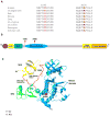
In silico analysis of the two ABL1 variants identified in this study. (A) The Tyr245 and Ala356 residues are conserved from human to zebrafish (prepared based on Ensembl browser genomic alignments). (B) The schematic view of ABL1 1b protein isoform and its domains. The Tyr245 residue localizes in the linker region between the SH2 and kinase domains, while the Ala356 localizes in the kinase domain. Prepared based on UniProt database domains, ID P00519. (C) The 3D structure of ABL1 1b protein isoform, its N-terminal domains, and the localization of the two mutated ABL1 residues. Based on the structural data from Nagar et al., 2003 (PDB ID 1OPL) using Swiss-PdbViewer.
Figure 4
Figure 4
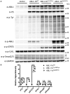
The effect of ABL1 variants (isoform 1b) on phosphorylation. (A) Overall phosphotyrosine levels and phosphorylation of specific ABL1 substrates were analyzed by transiently expressing the wildtype and mutant constructs in HEK 293T cells and immunoblotting. Both variants showed increased overall phosphotyrosine levels and phosphorylation of STAT5 when compared with wild-type. Increased phosphorylated ABL1 was observed for Ala356Thr but not Tyr245Cys due to the substitution of the Tyr245 residue, which is recognized by the anti-phospho-ABL1 antibody. No significant difference in the phosphorylation levels of CrKL, SMAD2 and SMAD3 between mutants and wild-type were observed. Antibodies used in the detection include anti-phosphotyrosine (p-Tyr) for the overall phosphotyrosine level, anti-phospho-ABL1 (p-ABL1), anti-phospho-STAT5 (p-STAT5), anti-phospho-CrkL (p-CrkL), and anti-phospho-SMAD2 and SMAD3 (p-Smad2/3) antibodies for the phosphorylation level of specific ABL1 substrates in the whole cell lysates. The level of GAPDH is used as an internal loading control. Experiments for each construct were performed in triplicates. Pound and asterisk symbols in the panel A denote p-Smad2 and p-Smad3 respectively. (B) Quantification of the Western blot results. Data are normalized to GAPDH protein levels, with the wild-type set at 1.0. **: P≤0.01; ***: P≤0.001; ****: P≤0.0001; n.s.: P>0.05.

References

    1. de Klein A, et al. A cellular oncogene is translocated to the Philadelphia chromosome in chronic myelocytic leukaemia. Nature. 1982;300:765–7. - PubMed
    1. Pye SM, et al. The effects of imatinib on pregnancy outcome. Blood. 2008;111:5505–8. - PMC - PubMed
    1. Apperley J. Issues of imatinib and pregnancy outcome. J Natl Compr Canc Netw. 2009;7:1050–8. - PubMed
    1. Jain N, Sharma D, Agrawal R, Jain A. A newborn with teratogenic effect of imatinib mesylate: a very rare case report. Med Princ Pract. 2015;24:291–3. - PMC - PubMed
    1. Ault P, et al. Pregnancy among patients with chronic myeloid leukemia treated with imatinib. J Clin Oncol. 2006;24:1204–8. - PubMed

MeSH terms

Supplementary concepts