. 2017 Apr;49(4):527-536.
doi: 10.1038/ng.3808. Epub 2017 Mar 13.

Disruption of the ATXN1-CIC complex causes a spectrum of neurobehavioral phenotypes in mice and humans

Affiliations

Disruption of the ATXN1-CIC complex causes a spectrum of neurobehavioral phenotypes in mice and humans

Hsiang-Chih Lu et al. Nat Genet. 2017 Apr.

Abstract

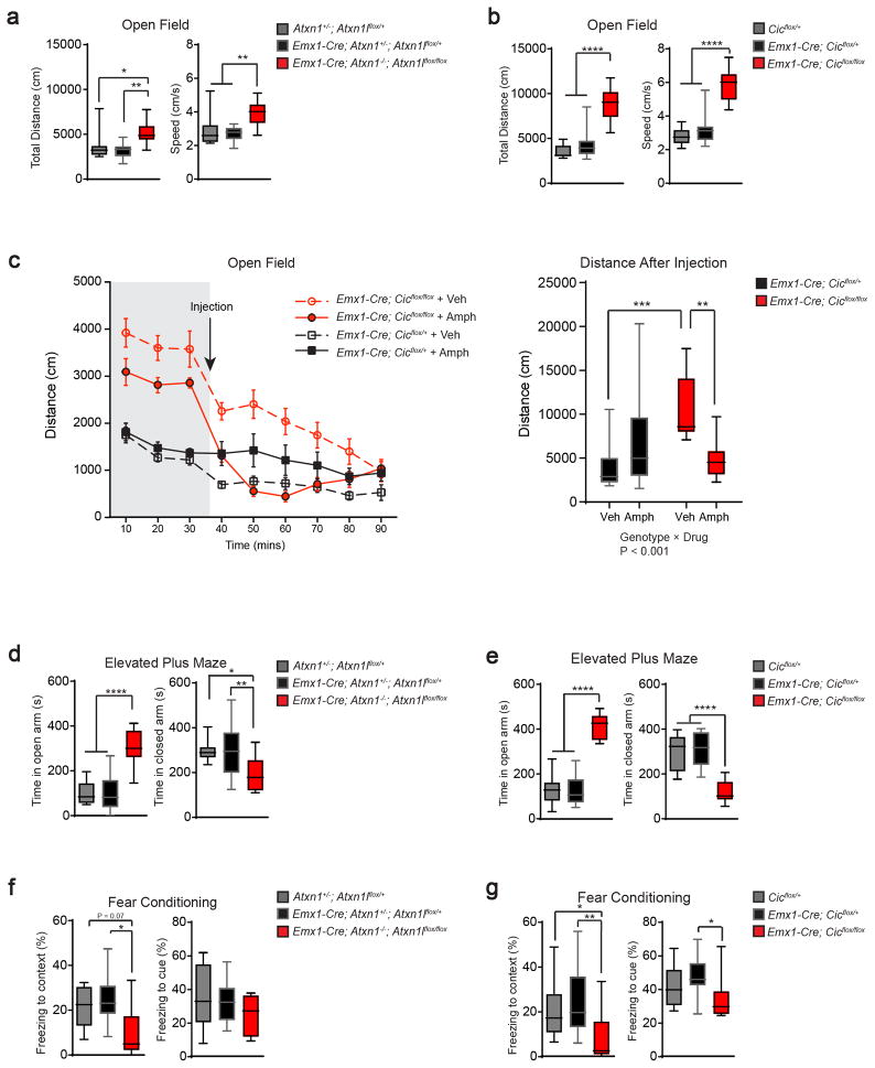
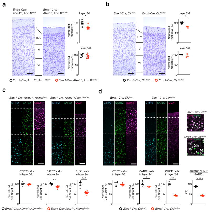
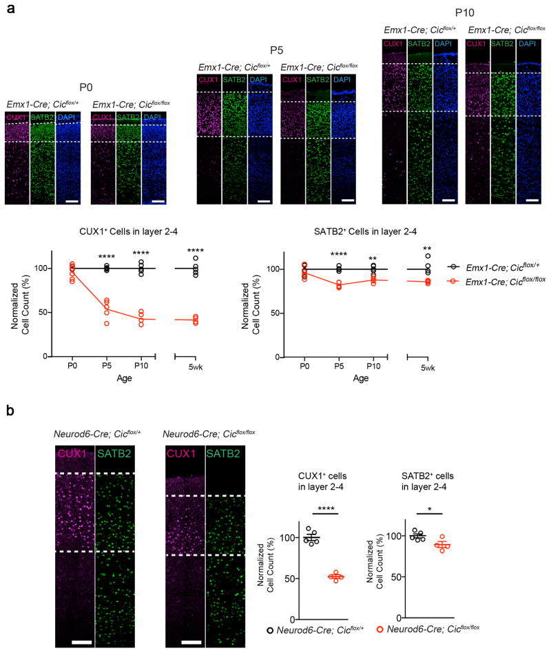
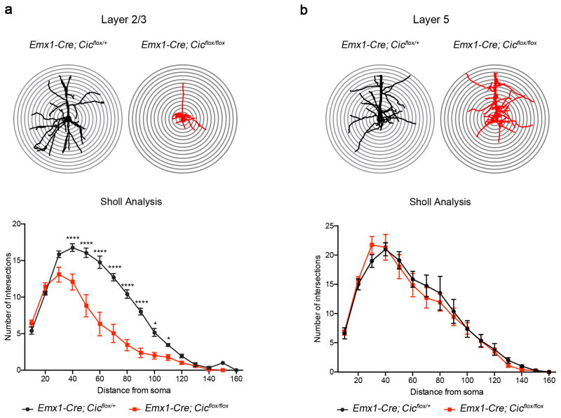
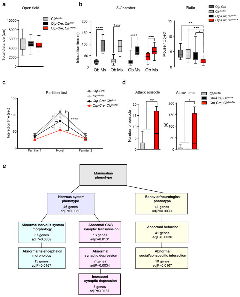
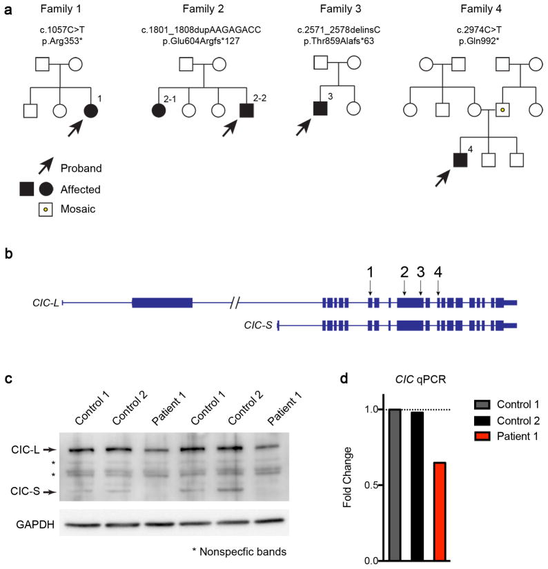
Gain-of-function mutations in some genes underlie neurodegenerative conditions, whereas loss-of-function mutations in the same genes have distinct phenotypes. This appears to be the case with the protein ataxin 1 (ATXN1), which forms a transcriptional repressor complex with capicua (CIC). Gain of function of the complex leads to neurodegeneration, but ATXN1-CIC is also essential for survival. We set out to understand the functions of the ATXN1-CIC complex in the developing forebrain and found that losing this complex results in hyperactivity, impaired learning and memory, and abnormal maturation and maintenance of upper-layer cortical neurons. We also found that CIC activity in the hypothalamus and medial amygdala modulates social interactions. Informed by these neurobehavioral features in mouse mutants, we identified five individuals with de novo heterozygous truncating mutations in CIC who share similar clinical features, including intellectual disability, attention deficit/hyperactivity disorder (ADHD), and autism spectrum disorder. Our study demonstrates that loss of ATXN1-CIC complexes causes a spectrum of neurobehavioral phenotypes.


Conflict of interest statement

Competing Financial Interests

None declared.

Figures

Figure 1
Figure 1. Deleting Atxn1-Atxn1l or Cic in the developing forebrain results in behavioral abnormalities
(a, b) In the open field test, both Atxn1-Atxn1l and Cic conditional knockout mice showed increased exploratory activities as demonstrated by increased total distance travelled and increased movement speed (n = 9–16, 12–13 weeks old). (c) After recording 30 minutes of baseline activity, we administered low-dose amphetamine (2 mg/kg body weight) and monitored locomotion for 60 minutes. Low-dose amphetamine calmed the Cic conditional knockout mice (n = 10–14, 12–13 weeks old). (d, e) In the elevated plus maze test, both mutant mice spent more time in the open arm and less time in the closed arm (n = 11–18, 12–13 weeks old). (f, g) Both mutant mouse lines had reduced freezing in the fear conditioning test (n = 8–13, 16–20 weeks old). Data are represented as mean ± s.e.m. in the left panel of 1c. All Other data are presented in box-and-whisker plots, with center lines represent median, box limits represent interquartile range, and whiskers represent minimum to maximum data range. *: P < 0.05; **: P < 0.01; ***: P < 0.001; ****: P < 0.0001
Figure 2
Figure 2. Deleting Atxn1-Atxn1l or Cic in the developing forebrain results in reduced thickness of upper cortical layers
(a, b) By Nissl staining, both mutant mouse lines had reduced thickness of layer 2–4, but not layer 5–6 (n = 3–5, scale bar = 100 μm). (c, d) Immunofluorescent staining of the cortex from 5-week old control and mutant animals. The number of CTIP2+ cells in layer 5–6 did not change, but the numbers of SATB2+ and CUX1+ cells were reduced in the mutant cortex (n = 4–5, scale bar = 100 μm). (d, inset) Mutant mice showed fewer layer 2–4 SATB2+ cells coexpressing CUX1. (Arrowhead: SATB2 single positive cells.) Data are represented as mean ± s.e.m. *: P < 0.05; **: P < 0.01; ***: P < 0.001; ****: P < 0.0001
Figure 3
Figure 3. The histological defects in Emx1-Cre Cic mutant animals occur postnatally in post-mitotic neurons
(a) The numbers of CUX1+ cells and SATB2+ cell in layer 2–4 diminished postnatally. Immunofluorescent staining with CUX1 and SATB2 revealed that the number of CUX1+ cells and SATB2+ cells at P0 were comparable among control and mutant animals. The number gradually decreased in the mutant mice, and by P10 the numbers were comparable to those from 5-week old animals (n = 4–6, scale bar = 100 μm). (The 5-week old data are from Figure 2.) (b) Immunofluorescent staining with CUX1 and SATB2 revealed that the numbers of layer 2–4 CUX1+ and SATB2+ cells were reduced in Neurod6-Cre conditional mutants, which removes Cic in post-mitotic neurons (n = 4–5, scale bar = 100 μm). Data are represented as mean ± s.e.m. *: P < 0.05; ****: P < 0.0001
Figure 4
Figure 4. Morphological defects in layer 2/3 pyramidal neurons in the Emx1-Cre Cic conditional knockout mice
(a) Sholl analysis showed that mutant layer 2/3 pyramidal neurons had reduced dendritic complexity as compared with control animals. (b) The dendritic complexity in layer 5 neurons was not affected in mutant mice (n = 4–6 animals, 2–4 cells per animal). Data are represented as mean ± s.e.m. *: P < 0.05; ****: P < 0.0001
Figure 5
Figure 5. Deleting Cic from the hypothalamus and medial amygdala results in abnormal social behavior
(a) The Otp-Cre conditional knockout mice showed normal exploratory activities in the open-field test (n = 16–28, 8–12 weeks old). (b) In the three-chamber test, the Otp-Cre conditional mutant animals still preferred mouse (Ms) over object (Ob), but they spent less time with mouse as compare to controls (n = 15–19, 8–12 weeks old). (c) In the partition test, the Otp-Cre; Cicflox/flox mice spent less time interacting with the novel partner mice. The Otp-Cre; Cicflox/+ mice had an intermediate phenotype (n = 14–19, 8–12 weeks old). (d) The Otp-Cre conditional knockout males were more aggressive in the resident-intruder test than controls, showing more and longer attacks toward a male intruder (n = 9–11, 8–12 weeks old). (e) Gene set enrichment analysis of differentially expressed genes between Otp-Cre; Cicflox/flox; ROSAfsTRAP and Otp-Cre; ROSAfsTRAP neurons. Data are represented as mean ± s.e.m. in c. All other data are presented in box-and-whisker plots, with center lines represent median, box limits represent interquartile range, and whiskers represent minimum to maximum data range. *: P < 0.05; **: P < 0.01; ***: P < 0.001; ****: P < 0.0001
Figure 6
Figure 6. Identification of heterozygous CIC truncating mutations in four families
(a) Pedigrees of four families with CIC truncating mutations. Both patients in the second family have the same mutation, but neither parent harbors the mutation in their somatic DNA; one of the parents is presumed to be germline mosaic. Sanger sequencing of the CIC variant regions in the two unaffected siblings revealed they do not carry the same mutations in CIC. The father in the fourth family is a low-grade somatic mosaic as demonstrated by Sanger sequencing. (b) Genomic locus of human CIC showing the location of the mutations found in the four families. All four mutations are predicted to create premature stop codons. (c) Western blot analysis of fibroblasts from patient 1 and two controls showing that CIC protein levels were reduced in the patient. Images were cropped for better presentation. (d) qPCR of CIC from patient 1 and control fibroblasts showed that the RNA levels of CIC were reduced in fibroblasts from the patient. The expression levels were normalized to control 1, and fold changes were plotted. Five to six technical repeats were performed for each sample.

References

    1. Orr HT, et al. Expansion of an unstable trinucleotide CAG repeat in spinocerebellar ataxia type 1. Nat Genet. 1993;4:221–6. - PubMed
    1. Burright EN, et al. SCA1 transgenic mice: a model for neurodegeneration caused by an expanded CAG trinucleotide repeat. Cell. 1995;82:937–48. - PubMed
    1. Matilla A, et al. Mice lacking ataxin-1 display learning deficits and decreased hippocampal paired-pulse facilitation. J Neurosci. 1998;18:5508–16. - PMC - PubMed
    1. Watase K, et al. A long CAG repeat in the mouse Sca1 locus replicates SCA1 features and reveals the impact of protein solubility on selective neurodegeneration. Neuron. 2002;34:905–19. - PubMed
    1. Asher M, Johnson A, Zecevic B, Pease D, Cvetanovic M. Ataxin-1 regulates proliferation of hippocampal neural precursors. Neuroscience. 2016 - PubMed