. 2017 Mar 13;6(3):e004756.
doi: 10.1161/JAHA.116.004756.

Activation of CD137 Signaling Promotes Angiogenesis in Atherosclerosis via Modulating Endothelial Smad1/5-NFATc1 Pathway

Affiliations

Activation of CD137 Signaling Promotes Angiogenesis in Atherosclerosis via Modulating Endothelial Smad1/5-NFATc1 Pathway

Jiayi Weng et al. J Am Heart Assoc. .

Retraction in

Abstract

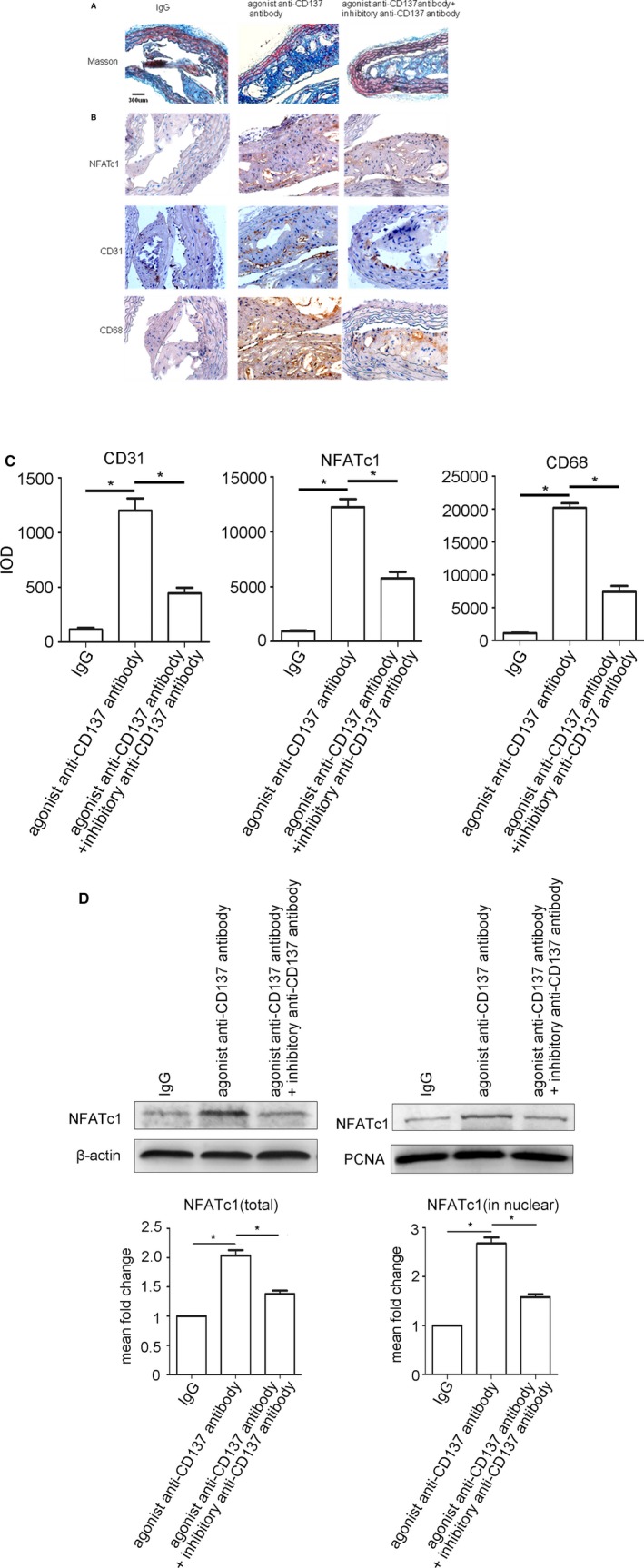
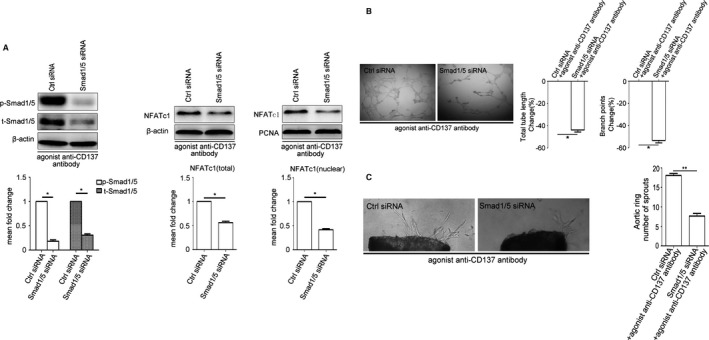
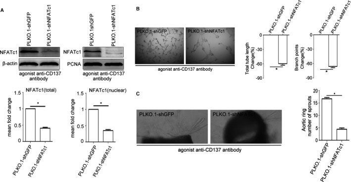
Background: Excessive angiogenesis is a key feature of vulnerable atherosclerotic plaques, and is considered an independent predictor of cardiovascular risk. CD137 signaling has previously been shown to be involved in atherosclerosis. However, the possible role of CD137 signaling in regulating angiogenesis has not been reported.

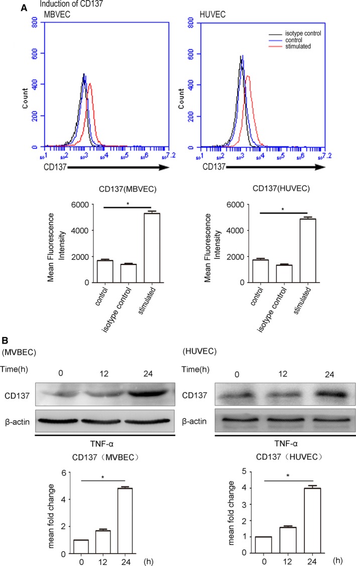
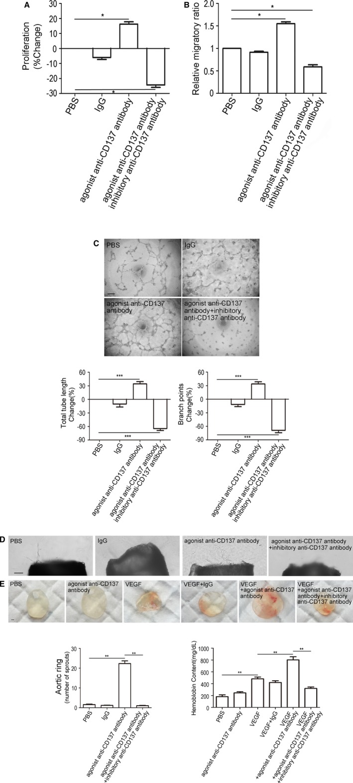
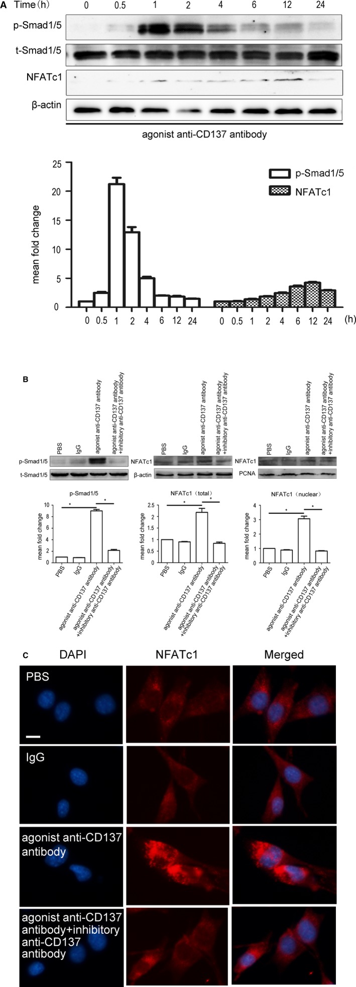
Methods and results: Apolipoprotein E-deficient (ApoE-/-) mice were used as the in vivo model of atherosclerosis. Masson and immunohistochemical analysis of atherosclerotic plaques and Matrigel plug assay were used to evaluate the angiogenesis. Human umbilical vein endothelial cells and mouse brain microvascular endothelial cells were used as in vitro and ex vivo models to study how CD137 signaling affects angiogenesis. Matrigel tube formation assay, mouse aortic ring angiogenesis assay, and migration and proliferation assay were employed to assess angiogenesis. Western blot was used to detect protein expression. We found increased neovessel formation in atherosclerotic plaques of ApoE-/- mice treated with agonist anti-CD137 antibody. Activation of CD137 signaling induced angiogenesis, endothelial proliferation, and endothelial cell migration. CD137 signaling activates the pro-angiogenic Smad1/5 pathway, induces the phosphorylation of Smad1/5 and nuclear translocation of p-Smad1/5, which in turn promotes the expression and translocation of NFATc1. Blocking CD137 signaling with inhibitory anti-CD137 antibody could inhibit this activation and attenuated agonist anti-CD137 antibody-induced angiogenesis.

Conclusions: These findings suggest that CD137 signaling is a new regulator of angiogenesis by modulating the Smad1/5-NFATc1 pathway.

Keywords: CD137; Smad1/5; angiogenesis; atherosclerosis; nuclear factor of activated T cells 1.


Figures

Figure 1
Figure 1
CD137 signaling induces NFATc1 expression and contributes to angiogenesis in atherosclerotic plaques in ApoE−/− mice. A, Masson's trichrome was performed to distinguish collagenous fiber (blue) and muscle fiber (red). Scale bar, 300 μm. B, Immunostaining of cross‐sections of the atherosclerotic plaques in ApoE−/− mice (NFATc1, CD31, CD68, respectively) as well as their quantitative analysis (C) CD31: IgG group median=104, IQR=58, n=5, agonist anti‐CD137 antibody group median=1103, IQR=339, n=5, agonist+inhibitory anti‐CD137 antibody group median=439, IQR=185, n=5. NFATc1: IgG group median=856, IQR=394, n=5, agonist anti‐CD137 antibody group median=12 116, IQR=2462, n=5, agonist+inhibitory anti‐CD137 antibody group median=5537, IQR=1651, n=5. CD68: IgG group median=958, IQR=350, n=5, agonist anti‐CD137 antibody group median=19 830, IQR=3090, n=5, agonist+inhibitory anti‐CD137 antibody group median=7694, IQR=2432, n=5. D, Western blot analysis of total and nuclear proteins levels of NFATc1 isolated from the atherosclerotic plaques in ApoE−/− mice. Average densitometric values normalized against those of internal control from three independent experiments are shown in the bar graph. NFATc1 (total): agonist anti‐CD137 antibody group median=1.98, IQR=0.13, n=5, agonist+inhibitory anti‐CD137 antibody group median=1.33, IQR=0.12, n=5. NFATc1 (in nuclear): agonist anti‐CD137 antibody group median=2.67, IQR=0.42, n=5, agonist+inhibitory anti‐CD137 antibody group median=1.59, IQR=0.21, n=5. *P<0.05. IgG indicates immunoglobulin G; IQR, interquartile range; NFATc1, nuclear factor of activated T cells 1.
Figure 2
Figure 2
TNF‐α stimulates CD137 expression in ECs. A, CD137 expressed on MBVECs and HUVECs after 24 hours of incubation with (red line) or without (blue) TNF‐α at 10 ng/mL. The black line represents antibody isotype‐matched control IgG. The statistical data analyses from 3 separate experiments are shown as bar graphs. MBVECs: control median=1753, IQR=321, n=5, isotype control median=1392, IQR=275, n=5, stimulated median=5297, IQR=629, n=5. HUVECs: control median=1724, IQR=429, n=5, isotype control median=1298, IQR=288, n=5, stimulated median=4753, IQR=484, n=5. B, The protein detected by Western blot in MBVEC and HUVECs after 24 hours of incubation with or without TNF‐α at 10 ng/mL. Average densitometric values normalized against those of internal control from 3 independent experiments are shown in the bar graph. MBVECs: isotype control median=1.67, IQR=0.29, n=5 stimulated median=4.84, IQR=0.34, n=5. HUVECs: isotype control median=1.57, IQR=0.24, stimulated median=3.83, IQR=0.35, n=5. *P<0.05. ECs indicates endothelial cells; HUVECs, human umbilical vein endothelial cells; IgG, immunoglobulin G; IQR, interquartile range; MBVEC, mouse brain microvascular endothelial cells; TNF‐α, tumor necrosis factor‐α.
Figure 3
Figure 3
CD137 signaling promotes angiogenesis in vivo, vitro, and ex vivo. A, The MBVEC treated with agonist anti‐CD137 antibody exhibit enhanced cell proliferation, and there is a reduction in inhibitory anti‐CD137 antibody‐treated cells. IgG group median=−5.7, IQR=4.97, n=5, agonist anti‐CD137 antibody group median=16.06, IQR=5.88, n=5, agonist+inhibitory anti‐CD137 antibody group median=−23.65, IQR=5.85, n=5. B, The MBVEC migration through a porous membrane (Transwell migration assay) over 24 hours was increased following addition of agonist anti‐CD137 antibody compared to control, and was inhibited by antiCD137 antibody. IgG group median=0.93, IQR=0.07, n=5, agonist anti‐CD137 antibody group median=1.55, IQR=0.14, n=5, agonist+inhibitory anti‐CD137 antibody group median=0.59, IQR=0.16, n=5. C, Increased HUVEC tube and branch formation after addition of agonist anti‐CD137 antibody, and inhibition by inhibitory anti‐CD137 antibody. Scale bar, 100 μm. Total tube length: IgG group median=−5.55, IQR=22.19, n=5, agonist anti‐CD137 antibody group median=36.85, IQR=18.28, n=5, agonist+inhibitory anti‐CD137 antibody group median=−64.55, IQR=17.3, n=5. Branch points: IgG group median=−8.38, IQR=17.65, n=5, agonist anti‐CD137 antibody group median=33.05, IQR=18.22, n=5, agonist+inhibitory anti‐CD137 antibody group median=−67.65, IQR=20.2, n=5. D, Vessel outgrowth in the aortic ring assay is enhanced by agonist anti‐CD137 antibody, and is attenuated by inhibitory anti‐CD137 antibody. Scale bar, 100 μm. PBS median=1.60, IQR=1.00, n=5, IgG group median=1.25, IQR=0.41, n=5, agonist anti‐CD137 antibody group median=22.06, IQR=2.63, n=5, agonist+inhibitory anti‐CD137 antibody group median=1.00, IQR=0.50, n=5. E, In Matrigel plug assay, Matrigel containing PBS, VEGF (100 ng/mL), agonist anti‐CD137 antibody (100 μg/mL), or inhibitory anti‐CD137 antibody (100 μg/mL) was injected subcutaneously into C57BL/6J mice. Ten days later, the Matrigel plugs were excised and photographed using a camera. Red color indicates abundant red blood cells. Hemoglobin content of Matrigel plugs from groups of mice was quantified by using QuantiChrom Hemoglobin Assay Kit. PBS median=186, IQR=58, n=5, agonist anti‐CD137 antibody group median=248.5, IQR=38.2, n=5, VEGF group median=479.0, IQR=53.3, n=5, VEGF+IgG group median=416.5, IQR=61.5, n=5. VEGF+agonist anti‐CD137 antibody group median=805.2, IQR=101.6, n=5, VEGF+agonist+inhibitory anti‐CD137 antibody group median=312.5, IQR=51.5, n=5. Scale bar, 1000 μm. All images shown are representative and values are expressed as mean±SEM. *P<0.05, **P<0.01, ***P<0.001. HUVECs indicates human umbilical vein endothelial cells; IgG, immunoglobulin G; IQR, interquartile range; MBVEC, mouse brain microvascular endothelial cells; PBS, phosphate‐buffered saline; VEGF, vascular endothelial growth factor.
Figure 4
Figure 4
CD137 signaling regulated phosphorylation of Smad1/5 and NFATc1 expression. A, Activation of CD137 signaling induces phosphorylation of Smad1/5 and expression of NFATc1 in MBVEC in a time‐dependent manner, and reached the peak at 1 and 12 hours, respectively. P‐Smad1/5: 0.5 hours median=2.43, IQR=0.62, 1 hour median=21.69, IQR=4.12, 2 hours median=12.58, IQR=3.30, 4 hours median=5.04, IQR=1.02, 6 hours median=1.99, IQR=0.18, 12 hours median=1.82, IQR=0.42, 24 hours median=1.49, IQR=0.21, n=3. NFATc1 0.5 hours median=0.90, IQR=0.26, 1 hour median=1.24, IQR=0.27, 2 hours median=1.75, IQR=0.21, 4 hours median=2.26, IQR=0.43, 6 hours median=3.32, IQR=0.58, 12 hours median=3.99, IQR=0.53, 24 hours median=2.81, IQR=0.25, n=3. B, MBVEC were treated with PBS, IgG (5 μg/mL), agonist anti‐CD137 antibody (5 μg/mL) or anti‐CD37 (5 μg/mL) for 1 or 12 hours. The protein levels of total and nuclear were determined by Western blot. Agonist anti‐CD137 antibody stimulates Smad1/5 phosphorylation (p‐Smad1/5) and NFATc1 expression, and this induction was inhibited by inhibitory anti‐CD137 antibody. Average densitometric values normalized against those of internal control from 3 independent experiments are shown in the bar graph. P‐Smad5/1 IgG group median=0.90, IQR=0.07, n=5, agonist anti‐CD137 antibody group median=9.13, IQR=0.89, n=5, agonist+inhibitory anti‐CD137 antibody group median=2.05, IQR=0.59, n=5. NFATc1 (total) IgG group median=0.90, IQR=0.05, n=5, agonist anti‐CD137 antibody group median=2.14, IQR=0.60, n=5, agonist+inhibitory anti‐CD137 antibody group median=0.83, IQR=0.19, n=5. NFATc1 (nuclear) IgG group median=0.89, IQR=0.09, n=5, agonist anti‐CD137 antibody group median=3.00, IQR=0.60, n=5, agonist+inhibitory anti‐CD137 antibody group median=0.81, IQR=0.08, n=5. *P<0.05. C, Immunofluorescence revealed that activation of CD137 signaling can induce NFATc1 expression and nuclear translocation. DAPI indicates 4′,6‐diamidino‐2‐phenylindole; IgG, immunoglobulin G; IQR, interquartile range; MBVEC, mouse brain microvascular endothelial cells; NFATc1, nuclear factor of activated T cells 1; PBS, phosphate‐buffered saline.
Figure 5
Figure 5
siRNASmad1/5 attenuates the expression of NFATc1 in HUVEC and angiogenesis in vitro induced by CD137 signaling. A, Western blot analysis revealed that siRNA knockdown of Smad1/5 in HUVEC results in reduced Smad1/5 phosphorylation and inhibits agonist anti‐CD137 antibody induced NFATc1 expression and nuclear translocation. Average densitometric values normalized against those of internal control from 3 independent experiments are shown in the bar graph. P‐Smad1/5 median=0.16, IQR=0.10, t‐Smad1/5 median=0.31, IQR=0.10, NFATc1 (total) median=0.60, IQR=0.12, NFATc1 (nuclear) median=0.46, IQR=0.10, n=3. B and C, Knockdown of Smad1/5 reduced agonist anti‐CD137 antibody‐mediated HUVEC Matrigel tube formation and decreased agonist anti‐CD137 antibody‐induced vessel outgrowth in aortic ring assays. Tube length: Smad1/5 siNRA median=−43.45, IQR=7.02, n=5. Branch points: Smad1/5 siNRA median=−54.00, IQR=7.92, n=5. Aortic ring ctrl siRNA median=17.60, IQR=1.20, Smad1/5 siNRA median=7.70, IQR=1.40, n=5. All images shown are representative and values are expressed as mean±SEM. *P<0.05, **P<0.01. Ctrl indicates control; HUVECs, human umbilical vein endothelial cells; IQR, interquartile range; NFATc1, nuclear factor of activated T cells 1; P‐Smad1/5, phosphorylated‐Smad1/5; SiRNA, small interfering RNA; t‐Smad1/5, total‐Smad1/5.
Figure 6
Figure 6
NFATc1 gene suppressing inhibited angiogenesis induced by CD137 signaling in vitro. A, NFATc1 knockdown stable cell lines (PLKO.1‐shNFATc1, PLKO.1‐shGFP as control) were treated with agonist anti‐CD137 antibody. Western blot reveals the protein level of NFATc1 in stable cell lines. Average densitometric values normalized against those of internal control from 3 independent experiments are shown in the bar graph. NFATc1 (total) median=0.40, IQR=0.09, NFATc1 (nuclear) median=0.36, IQR=0.11, n=3. B and C, Suppression of NFATc1 reduced agonist anti‐CD137 antibody‐mediated HUVEC Matrigel tube formation and decreased agonist anti‐CD137 antibody‐induced vessel outgrowth in aortic ring assays. Tube length: PLKO.1‐shNFATc1 median=−58.23, IQR=5.94, n=5. Branch points: PLKO.1‐shNFATc1 median=−66.83, IQR=5.77, n=5. Aortic ring PLKO.1‐shGFP median=16.5, IQR=1.93, PLKO.1‐shNFATc1 median=4.33, IQR=1.18, n=5. All images shown are representative and values are expressed as mean±SEM *P<0.05. HUVEC indicates human umbilical vein endothelial cell; IQR, interquartile range; NFATc1, nuclear factor of activated T cells 1; PCNA, proliferating cell nuclear antigen.; PLKO.1‐shGFP, PLKO.1‐short hairpin GFP; PLKO.1‐shNFATc1, PLKO.1‐short hairpin NFATc1

References

    1. Milei J, Parodi JC, Alonso GF, Barone A, Grana D, Matturri L. Carotid rupture and intraplaque hemorrhage: immunophenotype and role of cells involved. Am Heart J. 1998;136:1096–1105. - PubMed
    1. Moreno PR, Purushothaman KR, Fuster V, Echeverri D, Truszczynska H, Sharma SK, Badimon JJ, O'Connor WN. Plaque neovascularization is increased in ruptured atherosclerotic lesions of human aorta: implications for plaque vulnerability. Circulation. 2004;110:2032–2038. - PubMed
    1. Olofsson PS, Söderström LA, Wågsäter D, Sheikine Y, Ocaya P, Lang F, Rabu C, Chen L, Rudling M, Aukrust P, Hedin U, Paulsson‐Berne G, Sirsjö A, Hansson GK. CD137 is expressed in human atherosclerosis and promotes development of plaque inflammation in hypercholesterolemic mice. Circulation. 2008;117:1292–1301. - PubMed
    1. Jeon HJ, Choi JH, Jung IH, Park JG, Lee MR, Lee MN, Kim B, Yoo JY, Jeong SJ, Kim DY, Park JE, Park HY, Kwack K, Choi BK, Kwon BS, Oh GT. CD137 (4‐1BB) deficiency reduces atherosclerosis in hyperlipidemic mice. Circulation. 2010;121:1124–1133. - PubMed
    1. Yan J, Gong J, Liu P, Wang C, Chen G. Positive correlation between CD137 expression and complex stenosis morphology in patients with acute coronary syndromes. Clin Chim Acta. 2011;412:993–998. - PubMed

Publication types

MeSH terms

LinkOut - more resources