. 2017 Mar 13;6(3):e005328.
doi: 10.1161/JAHA.116.005328.

Mff-Dependent Mitochondrial Fission Contributes to the Pathogenesis of Cardiac Microvasculature Ischemia/Reperfusion Injury via Induction of mROS-Mediated Cardiolipin Oxidation and HK2/VDAC1 Disassociation-Involved mPTP Opening

Affiliations

Mff-Dependent Mitochondrial Fission Contributes to the Pathogenesis of Cardiac Microvasculature Ischemia/Reperfusion Injury via Induction of mROS-Mediated Cardiolipin Oxidation and HK2/VDAC1 Disassociation-Involved mPTP Opening

Hao Zhou et al. J Am Heart Assoc. .

Abstract

Background: The cardiac microvascular system ischemia/reperfusion injury following percutaneous coronary intervention is a clinical thorny problem. This study explores the mechanisms by which ischemia/reperfusion injury induces cardiac microcirculation collapse.

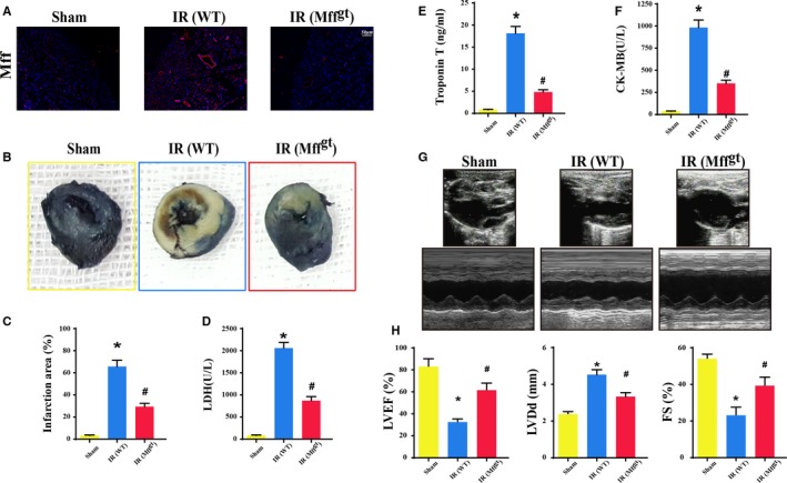
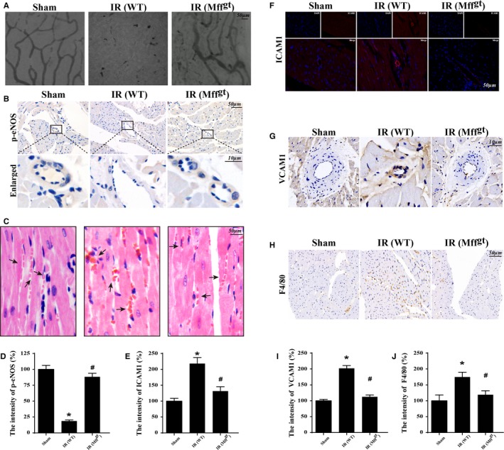
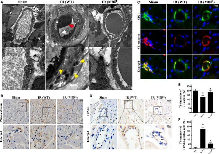
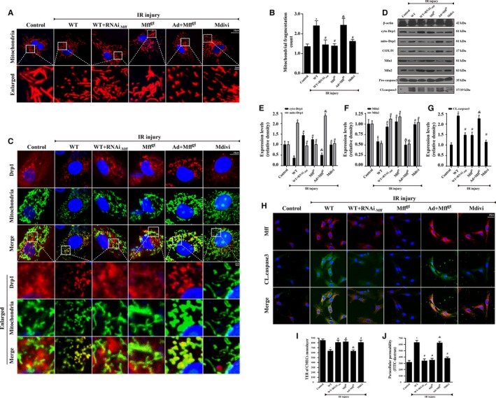
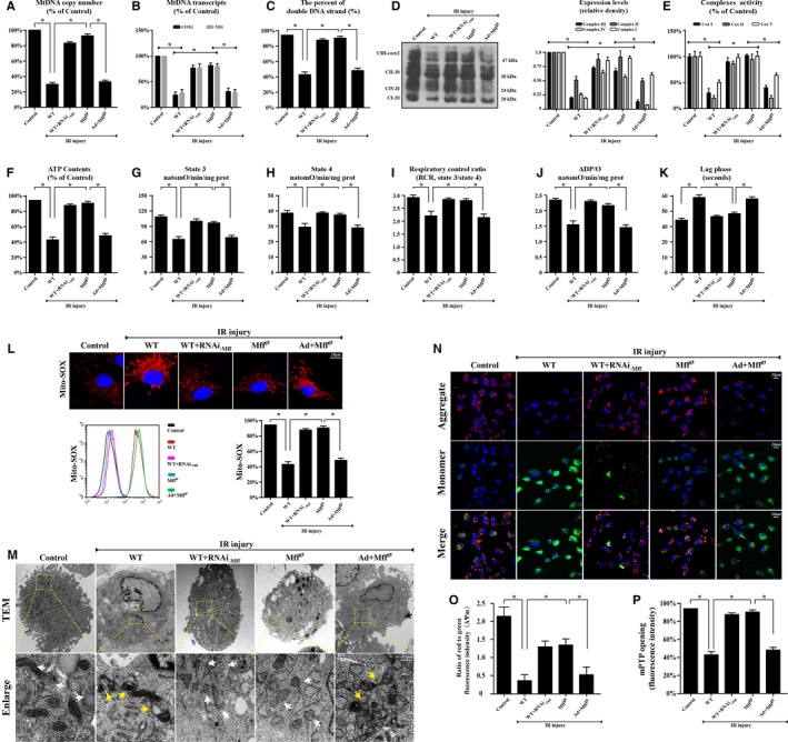
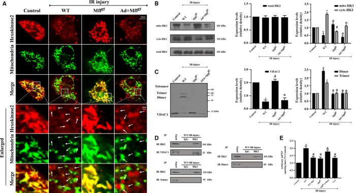
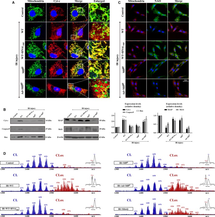
Methods and results: In wild-type mice, mitochondrial fission factor (Mff) expression increased in response to acute microvascular ischemia/reperfusion injury. Compared with wild-type mice, homozygous Mff-deficient (Mffgt) mice exhibited a smaller infarcted area, restored cardiac function, improved blood flow, and reduced microcirculation perfusion defects. Histopathology analysis demonstrated that cardiac microcirculation endothelial cells (CMECs) in Mffgt mice had an intact endothelial barrier, recovered phospho-endothelial nitric oxide synthase production, opened lumen, undivided mitochondrial structures, and less CMEC death. In vitro, Mff-deficient CMECs (derived from Mffgt mice or Mff small interfering RNA-treated) demonstrated less mitochondrial fission and mitochondrial-dependent apoptosis compared with cells derived from wild-type mice. The loss of Mff inhibited mitochondrial permeability transition pore opening via blocking the oligomerization of voltage-dependent anion channel 1 and subsequent hexokinase 2 separation from mitochondria. Moreover, Mff deficiency reduced the cyt-c leakage into the cytoplasm by alleviating cardiolipin oxidation resulting from damage to the electron transport chain complexes and mitochondrial reactive oxygen species overproduction.

Conclusions: This evidence clearly illustrates that microcirculatory ischemia/reperfusion injury can be attributed to Mff-dependent mitochondrial fission via voltage-dependent anion channel 1/hexokinase 2-mediated mitochondrial permeability transition pore opening and mitochondrial reactive oxygen species/cardiolipin involved cyt-c release.

Keywords: apoptosis; endothelial cell; ischemia/reperfusion injury; mitochondria.


Figures

Figure 1
Figure 1
Mitochondrial fission factor (Mff) contributed to infarct size expansion and cardiac function deterioration following ischemia/reperfusion (IR) injury in vivo, which was performed by 30 minutes of ischemia followed by 2 hours of reperfusion (n=6/group). A, Upregulation of Mff in microvascular in response to IR injury by immunofluorescence. B, Representative pictures of heart sections with 2,3,5‐triphenyltetrazolium chloride and Evans Blue staining. C, Bar graph indicates the infarct size expressed as a percentage of the total left ventricular area. D through F, The lactate dehydrogenase (LDH) release, Troponin T contents, and creatine kinase‐MB (CK‐MB) values were assessed via ELISA. G, Representative M‐mode echocardiography was performed after 2 hours of reperfusion with the parasternal long‐axis views in each group. H, Quantitative analysis of cardiac function by echocardiography. *P<0.05 vs the sham group; # P<0.05 vs the wild‐type (WT) group. FS indicates fractional shortening; LVDd, left ventricular diastolic dimension; LVEF, left ventricular ejection fraction; Mffgt, homozygous Mff‐deficient mice.
Figure 2
Figure 2
Mitochondrial fission factor (Mff) was involved in acute microcirculation malfunction during ischemia/reperfusion (IR) injury. A, Microvascular image detection by ink staining. B and D, Immunohistochemistry of phosphoendothelial nitric oxide synthase (p‐eNOS) expression. C, Hematoxylin and eosin staining for red blood cell morphology in different groups. E and F, The change of intercellular adhesion molecule–1 (ICAM1) expression in response to IR with or without Mff. G and I, Reduced vascular cell adhesion molecule–1 (VCAM1) contents under IR unless loss of Mff. H and J, Representative images of the accumulation of F4/80+ in myocardial tissue. *P<0.05 vs the sham group; # P<0.05 vs the wild‐type (WT) group. Mffgt indicates homozygous Mff‐deficient mice.
Figure 3
Figure 3
Loss of mitochondrial fission factor (Mff) maintained microvessels lumen patency, preserved endothelial barrier integrity, reduced vascular permeability, sustained cardiac microcirculation endothelial cell (CMEC) mitochondrial structure, and alleviated cellular death. A, Transmission electron microscopy was used to observe the structural changes of microvessel in response to ischemia/reperfusion (IR) injury, including microvascular wall destruction (white arrow), luminal stenosis (red arrow), and mitochondrial damage (yellow arrow) of CMEC. B, The leakage of plasma albumin out of the surface of the vessel wall into interstitial spaces suggested the increased microvascular permeability in response to after IR injury. C and E, The endothelial barrier integrity was assessed via vascular endothelial cadherin (VE‐cadherin) staining. Discontinuous punctiform or linear expression of VE‐cadherin could be observed in the IR injury group indicative of the broken endothelial barrier. However, loss of Mff could reverse the continuous linear of VE‐cadherin fluorescence. D and F, terminal deoxynucleotidyl transferase dUTP nick‐end labeling (TUNEL) assay to assess microvascular apoptosis. *P<0.05 vs the sham group; # P<0.05 vs the wild‐type (WT) group. Mffgt indicates homozygous Mff‐deficient mice; WT, wild‐type.
Figure 4
Figure 4
Mitochondrial fission factor (Mff) induced cardiac microcirculation endothelial cells (CMECs) apoptosis via excessive mitochondrial fission in vitro. Wild‐type (WT) mice– and homozygous Mff–deficient (Mffgt) mice–derived CMECs were named WT and Mffgt groups, respectively. Furthermore, Mff gain‐of‐function experiments were performed in CMECs from Mffgt using adenovirus vector (Ad+Mffgt group). Meanwhile, Mdivi‐1, an inhibitor of fission, was used in CMECs from WT mice as the negative control group. The ischemia/reperfusion (IR) injury in vitro was mimicked by 30 minutes of hypoxia with serum starvation and 2 hours of reoxygeneration. A and B, Mitochondria of CMECs are labeled with anti‐Tom20 antibody to determine the number of cells with mitochondria fragmentation. The boxed area under each micrograph is enlarged to determine mitochondria fragmentation. To assess changes in mitochondrial morphology quantitatively, the aspect ratio (AR; the mitochondrial length) and form factor (FF; the degree of mitochondrial branching) were calculated for each cell (the minimum value for both parameters is 1). High FF and AR values show healthy mitochondria, whereas low FF and AR indicate fragmented mitochondria. C, Co‐localization of dynamin‐related protein 1 (Drp1) and mitochondria. The boxed area under each micrograph represents the amplification of the white square. More Drp1 was located on fragmented mitochondria while loss of Mff could reduce Drp1 migration on mitochondria. D through F, IR increased mitochondria‐Drp1 expression. Meanwhile, IR also reduced proteins related to mitochondrial fusion. The control of cytoplasm and mitochondrial fractionation in the Western blots are β‐actin and cytochrome c oxidase subunit IV, respectively. D and G, Caspase‐3 activation (CL.caspase3 expression) was detected by Western blots. H, Mff and CL.caspase3 co‐location by immunofluorescence. I, Transendothelial electrical resistance (TER) and permeability examination in CMECs subjected to IR injury. Confluence of CMECs monolayer was assessed as stabilized basal resistance of 800 Ω. TER increases when endothelial cells adhere and spread out, and decreases when endothelial cells retract or lose adhesion, which is the marker of endothelial barrier function. J, Fluorescein isothiocyanate (FITC)–dextran clearance was measured to assess changes in endothelial permeability. FITC‐dextran was added on top of the inserts, allowing it to permeate through the cell monolayer. The increased endothelial permeability could retain more FITC‐dextran. Thus, the FITC content remaining on the plate after IR injury indicated the extent of permeability of CMECs. *P<0.05 vs the control group; # P<0.05 vs the WT group; & P<0.05 vs the Mffgt group.
Figure 5
Figure 5
Loss of mitochondrial fission factor (Mff) protected mitochondrial structure and function against ischemia/reperfusion (IR) injury. A, mtDNA copy number was assessed by complex IV segment. B, The transcript level of mtDNA was reflected by two different components: NADH dehydrogenase subunit 1 (ND1) encoded by the light chain of mtDNA and cytochrome c oxidase subunit I (COX I) encoded by the heavy chain of mtDNA. C, The percentage of double‐stranded mtDNA indicated the mtDNA strand breaks. D, The expression of mitochondrial electron transport chain complexes (ETCx). Complex III subunit core 2, 47 kDa—CIII‐core2”; complex II‐FeS subunit, 30 kDa—“CII‐30”; complex IV subunit II, 24 kDa—“CIVII”; complex I subunit NDUFB8, 20 kDa—“CI‐20”. E, Changes in ETCx I, II, and V activities measured spectrophotometrically. F, Change in ATP contents. G through K, Effect of Mff‐mediated fission on state 3 respiration, state 4 respiration, respiratory control ratio (RCR [state 3/state 4]), number of nmol ADP phosphorylated to atoms of oxygen consumed (ADP/O), and ADP phosphorylation lag phase (time elapsed in the depolarization/repolarization cycle during ADP phosphorylation). L, Mitochondrial reactive oxygen species contents. The curve chart indicates the quantitative flow cytometry results. M, Representative transmission electron microscopic (TEM) images of morphological changes in mitochondria in CMECs. White arrow: normal mitochondria that exhibit a spindle shape. Yellow arrow: the divisive mitochondria. N and O, Loss of Mff could preserve mitochondrial membrane potential (∆Ψm) by JC‐1 staining. P, Change in mitochondrial permeability transition pore (mPTP) opening. *P<0.05. Mffgt indicates homozygous Mff–deficient mice; WT, wild‐type.
Figure 6
Figure 6
Mitochondrial fission factor (Mff) deficiency inhibits mitochondrial permeability transition pore (mPTP) opening via the reduction of voltage‐dependent anion channel 1 (VDAC1) oligomerization and subsequent hexokinase 2 (HK2) separation from the mitochondria. A and B, The subcellular location of HK2 via immunofluorescence and Western blots. Ischemia/reperfusion (IR) injury contributes to the HK2 separation from the mitochondria to the cytoplasm, which is reversed by the loss of Mff. However, IR or Mff deficiency has no effects on the total content of HK2 but influences its subcellular distribution between the mitochondria and cytoplasm. C, The evaluation of VDAC1 oligomerization via ethylene glycolbis–based cross‐linking and immunoblotting using anti‐VDAC1 antibodies. IR injury increases VDAC1 oligomerization corresponding to molecular masses of 69 and 95 kDa, whereas the loss of Mff can reverse this change. D, HK2 and VDAC1 interaction assessed by immunoprecipitation (IP) experiments. E, Arbitrary mPTP opening time by tetramethylrhodamine ethyl ester fluorescence of diphenylamine‐2‐carboxilic acid (DpC) and 3‐bromopyruvate (3‐Brpa), which are the inhibitors of VDAC1 oligomerization and HK2 interaction, respectively. Arbitrary mPTP opening time was determined as the time when the tetramethylrhodamine ethyl ester fluorescence intensity decreased by half between the initial and residual fluorescence intensity. Cyclosporin A (CsA), an mPTP blocker, was used as the negative control. *P<0.05 vs the control group; # P<0.05 vs the wild‐type (WT) group; & P<0.05 vs the Mffgt group. The white arrow indicates the separated HK2 from mitochondria.
Figure 7
Figure 7
Mitochondrial fission factor (Mff)–mediated mitochondrial fission aggravates cyt‐c release via the oxidation of cardiolipin, which activates caspase‐9–involved mitochondrial death pathways. A, Immunostaining of cyt‐c leakage from mitochondria into cytoplasm. B, Changes in protein expression in association with mitochondrial apoptosis pathways. C, The changes in 10‐N‐nonyl acridine orange (NAO) fluorescence indicated ischemia/reperfusion (IR) injury induced cardiolipin (CL) oxidation. D, Assessment of molecular species of CL and its oxidation products. Left panel indicates the nonoxidized (blue) and the appearance of numerous oxidized (red) CL species after IR injury. Right inserts: 2‐dimensional chromatographic separation of nonoxidized and oxidized CL (CLox). IR injury oxidated CL via mitochondrial reactive oxygen species overproduction. *P<0.05 vs the control group; # P<0.05 vs the wild‐type (WT) group, & P<0.05 vs the homozygous Mff‐deficient (Mffgt) group.
Figure 8
Figure 8
Ischemia/reperfusion (IR)–mediated mitochondrial fission factor (Mff) upregulation is accompanied by increased mitochondrial fission and reduced fusion, leading to mitochondrial structural and function damage. Voltage‐dependent anion channel 1 (VDAC1) oligomerization in response to Mff‐mediated fission leads to the separation of hexokinase 2 (HK2) from the outer mitochondrial membrane due to the lower affinity between VDAC1 multimers and HK2, resulting in the opening of mitochondrial permeability transition pore (mPTP). Moreover, increased mitochondrial reactive oxygen species (mROS) induces cardiolipin (CL) peroxidation, which mediates cyt‐c release and activation of mitochondrial‐dependent apoptosis pathways. Eventually, the apoptosis of cardiac microcirculation endothelial cells (CMECs) contributed to microvascular perfusion defect, barrier damage, vascular wall destruction or luminal stenosis, increased vascular permeability, and phospho‐endothelial nitric oxide synthase (p‐eNOS) reduction that are responsible for acute cardiac microcirculatory IR injury.

References

    1. Kloner RA, Ganote CE, Jennings RB. The “no‐reflow” phenomenon after temporary coronary occlusion in the dog. J Clin Invest. 1974;54:1496–1508. - PMC - PubMed
    1. Dauber IM, VanBenthuysen KM, McMurtry IF, Wheeler GS, Lesnefsky EJ, Horwitz LD, Weil JV. Functional coronary microvascular injury evident as increased permeability due to brief ischemia and reperfusion. Circ Res. 1990;66:986–998. - PubMed
    1. Ito H. No‐reflow phenomenon and prognosis in patients with acute myocardial infarction. Nat Clin Pract Cardiovasc Med. 2006;3:499–506. - PubMed
    1. Weir RA, Murphy CA, Petrie CJ, Martin TN, Balmain S, Clements S, Steedman T, Wagner GS, Dargie HJ, McMurray JJ. Microvascular obstruction remains a portent of adverse remodeling in optimally treated patients with left ventricular systolic dysfunction after acute myocardial infarction. Circ Cardiovasc Imaging. 2010;3:360–367. - PubMed
    1. Dromparis P, Michelakis ED. Mitochondria in vascular health and disease. Annu Rev Physiol. 2013;75:95–126. - PubMed

MeSH terms

LinkOut - more resources