Review
. 2017 Oct 3;9(10):a028274.
doi: 10.1101/cshperspect.a028274.

Photoreceptor Cilia and Retinal Ciliopathies

Affiliations
Review

Photoreceptor Cilia and Retinal Ciliopathies

Kinga M Bujakowska et al. Cold Spring Harb Perspect Biol. .

Abstract

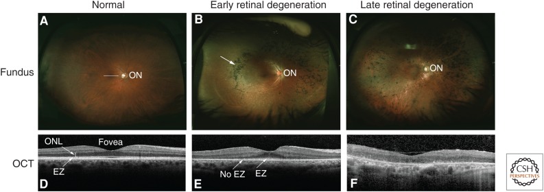
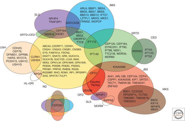
Photoreceptors are sensory neurons designed to convert light stimuli into neurological responses. This process, called phototransduction, takes place in the outer segments (OS) of rod and cone photoreceptors. OS are specialized sensory cilia, with analogous structures to those present in other nonmotile cilia. Deficient morphogenesis and/or dysfunction of photoreceptor sensory cilia (PSC) caused by mutations in a variety of photoreceptor-specific and common cilia genes can lead to inherited retinal degenerations (IRDs). IRDs can manifest as isolated retinal diseases or syndromic diseases. In this review, we describe the structure and composition of PSC and different forms of ciliopathies with retinal involvement. We review the genetics of the IRDs, which are monogenic disorders but genetically diverse with regard to causality.


Figures

Figure 1.
Figure 1.
Photoreceptor structure. Schematic representation of the rod photoreceptor with sensory cilia components indicated. The drawings to the left of the photoreceptor represent the cross-sectional view of the microtubule structure of the distal axoneme (Ax), proximal Ax, transition zone (TZ), and basal body (BB). RT, rootlet; OS, outer segment; CP, calyceal process; IS, inner segment; Ncl, nucleus; Syn, synapse.
Figure 2.
Figure 2.
Clinical features of inherited retinal degeneration (IRD). Fundus photos and optical coherence tomography (OCT) images of normal and diseased retinas are shown. (A) Wide-field fundus photo shows the appearance of a normal retina; the optic nerve (ON) is visible. (B) Fundus image from a patient with early retinal degeneration because of retinitis pigmentosa (RP). The arrow shows the pigment changes, which are characteristic of this disorder. (C) Fundus image from a patient with advanced retinal degeneration because of RP. (D) A cross-sectional image of the center of the normal retina (macula; white line in A) obtained with OCT shows normal retinal layers, including the outer nuclear layer (ONL), where photoreceptor cell nuclei are located. The central indentation is normal, and indicates the fovea. The white band showing the elipsoid zone (EZ, arrow) is generated from the junction of the inner and outer segments of photoreceptor cells, and its presence indicates normal photoreceptor cell and thus PSC structure. (E) The OCT image shows loss of photoreceptor cells peripherally, with the ONL visible only near the fovea. The EZ is evident centrally, but is lost more peripherally, indicating loss of PSCs by the more peripheral photoreceptor cells present. (F) The OCT image shows loss of the ONL and thus all of the photoreceptor cells in the macula of this patient consistent with greatly reduced central vision.
Figure 3.
Figure 3.
A Venn diagram showing a phenotypic and genetic overlap between different forms of ciliopathy. BBS, Bardet–Biedl syndrome; CED, cranioectodermal dysplasia (also known as Sensenbrenner syndrome); HL + SRI, hearing loss and sinorespiratory infections; JBS, Joubert syndrome; MKS, Meckel–Gruber syndrome; MORM, mental retardation, truncal obesity, retinal degeneration, and micropenis; OFD, oral-facial-digital syndrome; RD, retinal degeneration—nonsyndromic; SLS, Senior–Løken syndrome; SRTD, short-rib thoracic dysplasia; USH, Usher syndrome.

Similar articles

Cited by

References

    1. Abd El-Aziz MM, Barragan I, O’Driscoll CA, Goodstadt L, Prigmore E, Borrego S, Mena M, Pieras JI, El-Ashry MF, Safieh LA, et al. 2008. EYS, encoding an ortholog of Drosophila spacemaker, is mutated in autosomal recessive retinitis pigmentosa. Nat Genet 40: 1285–1287. - PMC - PubMed
    1. Abu-Safieh L, Al-Anazi S, Al-Abdi L, Hashem M, Alkuraya H, Alamr M, Sirelkhatim MO, Al-Hassnan Z, Alkuraya B, Mohamed JY, et al. 2012. In search of triallelism in Bardet–Biedl syndrome. Eur J Hum Genet 20: 420–427. - PMC - PubMed
    1. Ahmed ZM, Riazuddin S, Bernstein SL, Ahmed Z, Khan S, Griffith AJ, Morell RJ, Friedman TB, Wilcox ER. 2001. Mutations of the protocadherin gene PCDH15 cause Usher syndrome type 1F. Am J Hum Genet 69: 25–34. - PMC - PubMed
    1. Ahmed ZM, Kjellstrom S, Haywood-Watson RJL, Bush RA, Hampton LL, Battey JF, Riazuddin S, Frolenkov G, Sieving PA, Friedman TB. 2008. Double homozygous waltzer and Ames waltzer mice provide no evidence of retinal degeneration. Mol Vis 14: 2227–2236. - PMC - PubMed
    1. Akahori M, Tsunoda K, Miyake Y, Fukuda Y, Ishiura H, Tsuji S, Usui T, Hatase T, Nakamura M, Ohde H, et al. 2010. Dominant mutations in RP1L1 are responsible for occult macular dystrophy. Am J Hum Genet 87: 424–429. - PMC - PubMed