Review
. 2017 May 5;292(18):7295-7303.
doi: 10.1074/jbc.R117.777102. Epub 2017 Mar 13.

Contribution of type III interferons to antiviral immunity: location, location, location

Affiliations
Review

Contribution of type III interferons to antiviral immunity: location, location, location

Sergei V Kotenko et al. J Biol Chem. .

Abstract

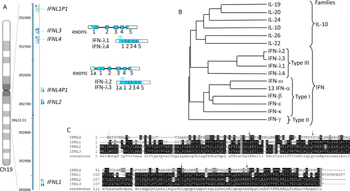
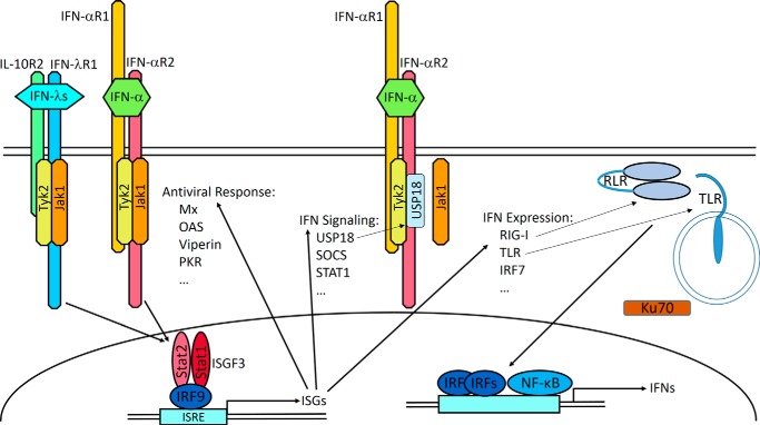
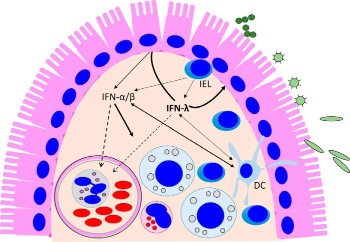
Type I interferons (IFN-α/β) and the more recently identified type III IFNs (IFN-λ) function as the first line of defense against virus infection and regulate the development of both innate and adaptive immune responses. Type III IFNs were originally identified as a novel ligand-receptor system acting in parallel with type I IFNs, but subsequent studies have provided increasing evidence for distinct roles for each IFN family. In addition to their compartmentalized antiviral actions, these two systems appear to have multiple levels of cross-regulation and act coordinately to achieve effective antimicrobial protection with minimal collateral damage to the host.

Keywords: antiviral agent; interferon; mucosal immunology; viral immunology; virology.


Conflict of interest statement

The content is solely the responsibility of the authors and does not necessarily represent the official views of the National Institutes of Health

Figures

Figure 1.
Figure 1.
Human type III IFNs. A, schematic representations of the chromosomal localization and intron/exon organization of the genes encoding human IFN-λs. The genes are transcribed in the direction indicated by the arrows. Genes encoding functional proteins are shown in blue and pseudogenes in green. Unspliced transcripts are schematically shown as strings of filled or open boxes (exons) joined by intervening solid lines (introns). Spliced transcripts are also shown as shaded/open boxes with vertical lines indicating the relative positions of former introns. The coding regions of exons are shaded, and the segments corresponding to the 5′- and 3′-untranslated regions are open (not shaded). The position of the signal peptide (SP) is shown in light blue. IFNL1 and IFNL4 genes are composed of five exons, whereas IFNL2 and IFNL3 genes have an additional upstream exon 1a. There are in-frame Met codons in both exons 1 and 1a; exon 1a extends the signal peptide by four additional amino acids. B, phylogenetic tree for IFN-λs with other IFNs and IL-10-related cytokines, (IL-10, IL-19, IL-22, IL-24, and IL-26) encoded in the human genome. Only one IFN-α was used in this alignment because the 13 human IFN-α subtypes have nearly identical sequences. C, sequence alignment of human IFN-λ proteins with the consensus sequence shown on the bottom. Arrows indicate positions of common introns (solid arrows) and of an additional intron, which is present only in IFNL2 and IFNL3 genes (dashed arrow).
Figure 2.
Figure 2.
IFN receptor complexes and signaling. IFN-λs and type I IFNs use distinct heterodimeric receptor complexes. The IFN-λs engage the unique IFN-λR1 and IL-10R2, a subunit also used by the IL-10, IL-22, and IL-26 receptor complexes. IFN-αR1 and IFN-αR2 form the active type I IFN receptor complex. IFN-λ binding to its receptor complex leads to the activation of receptor-associated JAK1 and Tyk2 kinases, which phosphorylate Tyr residues within the IFN-λR1 intracellular domain. These phosphotyrosine-based motifs serve as docking or recruitment sites for the latent transcriptional factors of the STAT family. The main STATs that become recruited and activated are STAT1 and STAT2, although activation of STAT3, STAT4, and STAT5 can be also detected (10, 19, 97). Phosphorylated STAT1 and STAT2 heterodimerize and interact with another transcription factor, IFN regulatory factor 9 (IRF9), leading to the formation of a transcription complex designated IFN-stimulated gene factor 3 (ISGF3). After translocation to the nucleus, ISGF3 binds to the IFN-specific response element (ISRE) that is commonly present in the promoter regions of hundreds of IFN-stimulated genes (ISGs), culminating in their transcriptional expression. The many ISGs encode a variety of antiviral mediators (98) enabling the establishment of an antiviral state effective against a broad spectrum of viruses. Subsets of ISGs encode proteins involved in virus recognition and IFN induction such as TLR, RIG-I-like receptors (RLR), and IRF7 as well as modulators of IFN signaling such as STAT1, USP18, and SOCS1 (99). USP18 binds to the IFN-αR2 intracellular domain, displaces JAK1, and suppresses IFN-α signaling. Ku70 is a DNA sensor that selectively triggers expression of IFN-λs (100).
Figure 3.
Figure 3.
IFN-λ-centric model on the production and action of IFNs at the epithelial barrier. IFN production in the intestine is induced by invading bacterial and viral pathogens and likely by the many microbial by-products present in the intestinal lumen. These stimuli trigger predominantly IFN-λ expression from epithelial cells, and these cells preferentially respond to type III IFNs. Therefore, it appears that the antiviral protection of IECs relies on the IFN-λ-based autocrine system. In addition to IECs, DCs present in the lamina propria beneath the epithelial surface produce and respond to both types of IFNs, but pDCs preferentially produce IFN-α. Intraepithelial T lymphocytes (IELs), which are in continuous contact with the epithelial layer, also secrete both type I and type III IFNs upon antigen stimulation (101). IFN-α/β secreted into the submucosa (solid arrows) act on lamina propria cells. Both IFN types can also act systemically through entry into the blood stream (dashed line).

Similar articles

Cited by

References

    1. Krause C. D., and Pestka S. (2015) Cut, copy, move, delete: the study of human interferon genes reveals multiple mechanisms underlying their evolution in amniotes. Cytokine 76, 480–495 - PubMed
    1. Qi Z., Nie P., Secombes C. J., and Zou J. (2010) Intron-containing type I and type III IFN coexist in amphibians: refuting the concept that a retroposition event gave rise to type I IFNs. J. Immunol. 184, 5038–5046 - PubMed
    1. Sang Y., Liu Q., Lee J., Ma W., McVey D. S., and Blecha F. (2016) Expansion of amphibian intronless interferons revises the paradigm for interferon evolution and functional diversity. Sci. Rep. 6, 29072. - PMC - PubMed
    1. Boudinot P., Langevin C., Secombes C. J., and Levraud J. P. (2016) The peculiar characteristics of fish type I interferons. Viruses 8, E298. - PMC - PubMed
    1. Kotenko S. V., and Langer J. A. (2004) Full house: 12 receptors for 27 cytokines. Int. Immunopharmacol. 4, 593–608 - PubMed

LinkOut - more resources