. 2017 Apr 21;292(16):6583-6599.
doi: 10.1074/jbc.M116.773150. Epub 2017 Mar 13.

Human cytomegalovirus-encoded viral cyclin-dependent kinase (v-CDK) UL97 phosphorylates and inactivates the retinoblastoma protein-related p107 and p130 proteins

Affiliations

Human cytomegalovirus-encoded viral cyclin-dependent kinase (v-CDK) UL97 phosphorylates and inactivates the retinoblastoma protein-related p107 and p130 proteins

Satoko Iwahori et al. J Biol Chem. .

Abstract

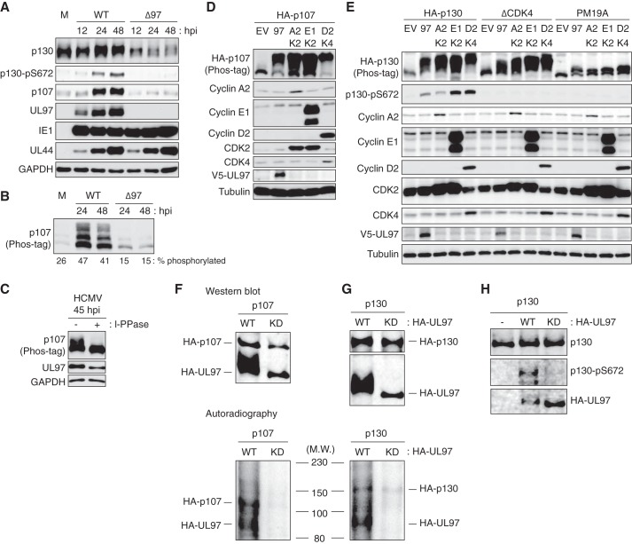
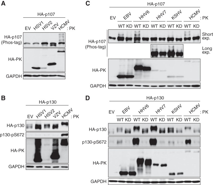
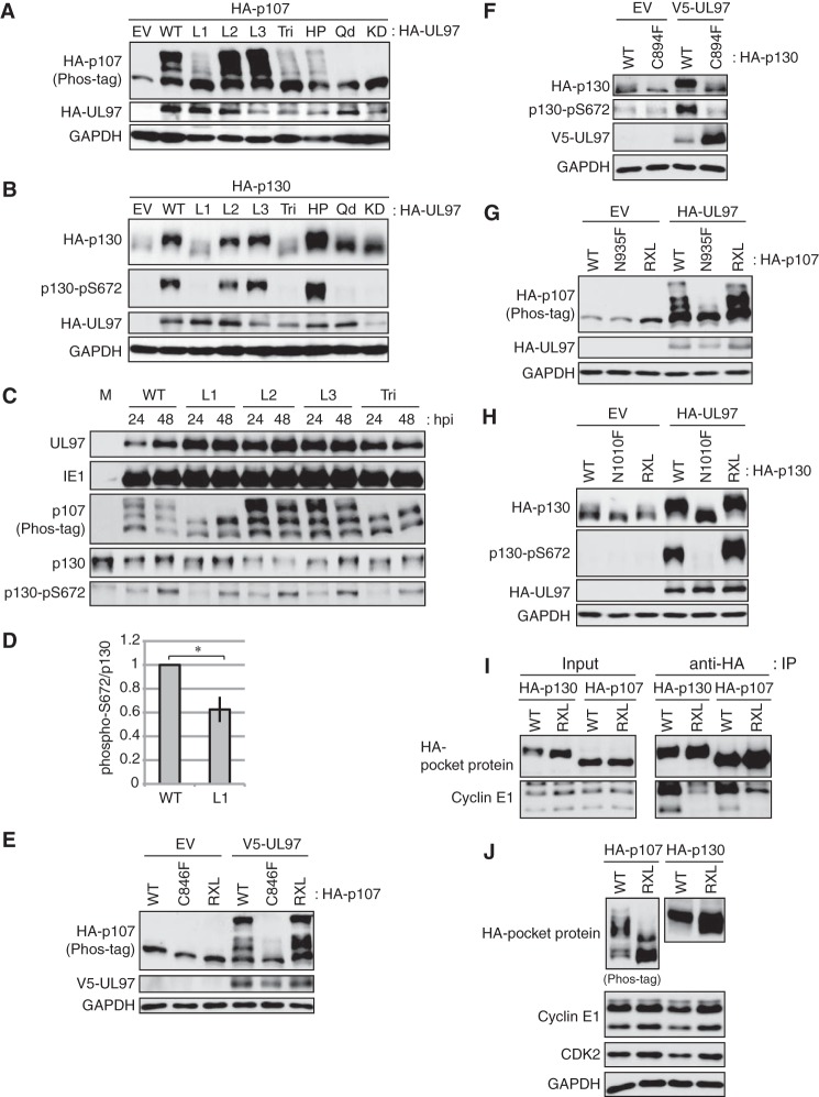
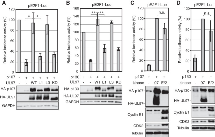
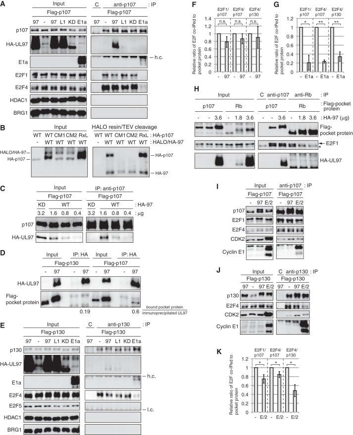
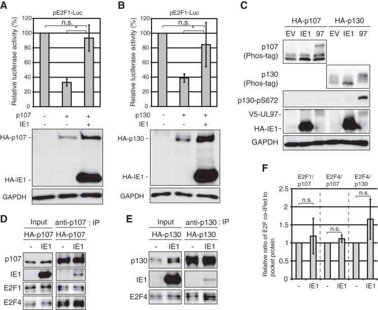
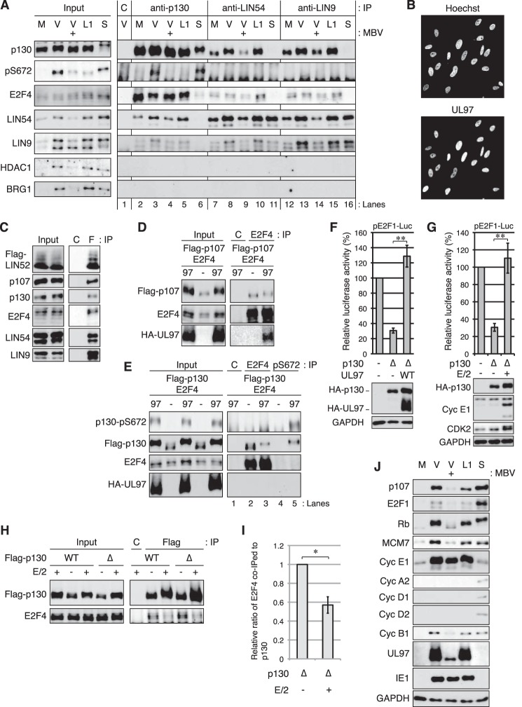
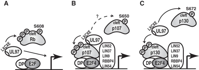
The human cytomegalovirus (HCMV)-encoded viral cyclin-dependent kinase (v-CDK) UL97 phosphorylates the retinoblastoma (Rb) tumor suppressor. Here, we identify the other Rb family members p107 and p130 as novel targets of UL97. UL97 phosphorylates p107 and p130 thereby inhibiting their ability to repress the E2F-responsive E2F1 promoter. As with Rb, this phosphorylation, and the rescue of E2F-responsive transcription, is dependent on the L1 LXCXE motif in UL97 and its interacting clefts on p107 and p130. Interestingly, UL97 does not induce the disruption of all p107-E2F or p130-E2F complexes, as it does to Rb-E2F complexes. UL97 strongly interacts with p107 but not Rb or p130. Thus the inhibitory mechanisms of UL97 for Rb family protein-mediated repression of E2F-responsive transcription appear to differ for each of the Rb family proteins. The immediate early 1 (IE1) protein of HCMV also rescues p107- and p130-mediated repression of E2F-responsive gene expression, but it does not induce their phosphorylation and does not disrupt p107-E2F or p130-E2F complexes. The unique regulation of Rb family proteins by HCMV UL97 and IE1 attests to the importance of modulating Rb family protein function in HCMV-infected cells.

Keywords: HCMV; cancer; cyclin-dependent kinase (CDK); herpesvirus; oncogene; phosphorylation; tumor suppressor gene.


Conflict of interest statement

The authors declare that they have no conflicts of interest with the contents of this article

Figures

Figure 1.
Figure 1.
UL97 phosphorylates p107 and p130 during HCMV lytic infection. HFFs were serum-starved for 48 h then infected with HCMV or UL97-null virus (Δ97) at an m.o.i. of 1. Whole-cell lysates were prepared at the indicated times postinfection, and equal amounts of protein from each sample were loaded onto a normal gel (A) or phosphate affinity (phos-tag) gel (B) and subjected to Western blotting analysis with the indicated antibodies. The percentage of phosphorylated p107 (compared with total) quantified by ImageJ is presented below the panel. M, mock infection. C, lysates from HCMV-infected HFFs were treated (+) or not (−) with λ protein phosphatase (l-PPase) and analyzed as in B. D, Saos-2 cells were transfected with expression plasmids for HA-tagged p107 (HA-p107) together with either an empty vector (EV), one expressing V5-tagged UL97 (97), cyclin A2 (A2)/CDK2 (K2), cyclin E1 (E1)/CDK2, or cyclin D2 (D2)/CDK4 (K4). Lysates harvested 48 h after transfection were analyzed by Western blotting with the indicated antibodies. E, transfections were performed as in D with HA-tagged p130 (HA-p130), p130ΔCDK4, or p130-PM19A. F and G, lysates of Saos-2 cells transfected with plasmids expressing HA-tagged p107 (HA-p107) or p130 (HA-p130) were immunoprecipitated with anti-p107 or p130 antibodies. Either wild-type (WT) or kinase-dead (KD) UL97 was incubated with immune-enriched p107 or p130 substrates in the presence of [γ-32P]ATP. Kinase reactions were resolved by SDS-PAGE and transferred to a nitrocellulose membrane. They were visualized by both autoradiography and Western blotting with an anti-HA antibody for detection of HA-UL97 and HA-p107 and an anti-p130 antibody for detection of HA-p130. M.W., molecular weight. H, kinase reaction was conducted as in F except normal ATP and bacterially purified GST-tagged p130 were utilized. Samples were analyzed by Western blotting with the indicated antibodies. Experiments were performed in biological triplicate except those in D and E that were performed in biological duplicates.
Figure 2.
Figure 2.
v-CDKs of EBV, HHV6, HHV7, and KSHV phosphorylate p107 and p130. Saos-2 cells were transfected with expression plasmids encoding HA-tagged p107 (A and C) or HA-tagged p130 (B and D) together with either an empty vector (EV) or an HA-tagged human herpesvirus protein kinase (HA-PK). Forty eight hours later, cells were harvested and subjected to Western blotting with the indicated antibodies. C, upper blot for HA-p107 corresponds to the blot after short exposure, and the bottom blot corresponds to the blot after long exposure. WT, wild type. KD, kinase-dead. The phosphorylation-dependent band shift of p107 was detected by phosphate affinity (phos-tag) gel electrophoresis. Experiments were performed in biological duplicate.
Figure 3.
Figure 3.
Phosphorylation of p107 and p130 by UL97 requires the L1 LXCXE motif of UL97 and the cleft of p107 and p130. A and B, Saos-2 cells were transfected with plasmids encoding HA-tagged p107 (A) or p130 (B) together with either an empty vector (EV), an expression plasmid for wild-type UL97 (WT), or an expression plasmid for UL97 alleles with the following mutations: L1, C151G; L2, C428G; L3, C693G; Tri, C151G/C428G/C693G; HP, W368A; quadruple (Qd), C151G/W368A/C428G/C693G; kinase-dead (KD), K355Q. After 48 h, cells were harvested and subjected to Western blotting with the indicated antibodies. C, serum-starved HFFs were infected with HCMV or the indicated LXCXE mutant viruses at an m.o.i. of 1. Whole-cell lysates were subjected to Western blotting analysis with the indicated antibodies. M, mock infection. D, level of p130 phosphorylated at Ser-672 normalized to total p130 was quantitated from wild-type or L1 mutant UL97 virus-infected cells at 48 h post-infection in experiments identical to those in C except at an m.o.i. of 2. Values are presented relative to the value in wild-type virus-infected cells (set at 1). Error bars denote the standard deviation. *, p < 0.05. E, Saos-2 cells were transfected with expression plasmids encoding V5-tagged wild-type UL97 together with either wild-type HA-tagged p107, a p107 cleft mutant (C846F), or a p107 RXL mutant. Forty eight hours later, lysates were harvested and subjected to Western blotting with the indicated antibodies. F, Saos-2 cells were transfected with expression plasmids encoding V5-tagged wild-type UL97 together with either wild-type HA-tagged p130 or a p130 cleft mutant (C894F). Forty eight hours later, lysates were harvested and subjected to Western blotting with the indicated antibodies. G, Saos-2 cells were transfected with expression plasmids encoding HA-tagged wild-type UL97 together with either wild-type HA-tagged p107, a p107 cleft mutant (N935F), or a p107 RXL mutant. Forty eight hours later, lysates were harvested and subjected to Western blotting with the indicated antibodies. H, Saos-2 cells were transfected with expression plasmids encoding HA-tagged wild-type UL97 together with either wild-type HA-tagged p130, a p130 cleft mutant (N1010F), or a p130 RXL mutant. Forty eight hours later, lysates were harvested and subjected to Western blotting with the indicated antibodies. I, Saos-2 cells were transfected with an expression plasmid for cyclin E1 together with HA-tagged wild-type or RXL mutant p107 or p130 expression plasmids. Lysates harvested 48 h after transfection were subjected to immunoprecipitation (IP) with the HA antibody. Input lysates and immunoprecipitates were analyzed by Western blotting with the indicated antibodies. J, Saos-2 cells were transfected with expression plasmids encoding cyclin E1 and CDK2 together with HA-tagged wild-type p107, a p107 RXL mutant, wild-type p130, or a p130 RXL mutant. Forty eight hours later, lysates were harvested and subjected to Western blotting with the indicated antibodies. The phosphorylation-dependent band shifts of both exogenously expressed and endogenous p107 were detected in phosphate affinity (phos-tag) gels. Experiments were performed in at least biological triplicates.
Figure 4.
Figure 4.
UL97 relieves p107- or p130-mediated repression of an E2F-responsive promoter. A, Saos-2 cells were transfected with a luciferase reporter driven by the E2F1 promoter together with either an empty vector (−) or an expression plasmid for HA-tagged p107, and either an empty vector or an expression plasmid for the indicated allele of UL97. Lysates harvested 48 h after transfection were analyzed for luciferase activity (top) and protein expression with the indicated antibodies (bottom). Luciferase activity was normalized to total protein concentration and is presented relative to the activity of the reporter without p107 or UL97 (set at 100%). B, luciferase and Western blotting analyses were performed as in A except with HA-tagged p130. Luciferase activity is presented relative to the activity of the reporter without p130 or UL97 (set at 100%). C, luciferase and Western blotting analyses were performed as in A except cyclin E1/CDK2 (E/2) was included. Luciferase activity is presented relative to the activity of the reporter with UL97 (97) (set at 100%). D, luciferase and Western blotting analyses were performed as in B except cyclin E1/CDK2 was included. Luciferase activity is presented relative to the activity of the reporter with UL97 (set at 100%). Error bars denote the standard deviation of more than three biological replicates. *, p < 0.05; **, p < 0.01; n.s., not significant.
Figure 5.
Figure 5.
Stable p130 knockdown reduces HCMV productive replication. A, equal amounts of protein lysates from subconfluent, serum-starved NHDFs transduced with a retrovirus expressing a scrambled shRNA sequence (Scr) or either of two shRNAs targeting p130 (130.1 or 130.3) were analyzed by Western blotting with the indicated antibodies. Dashed line indicates the removal of irrelevant lanes and merging of the remaining images. B, serum-starved Scr, 130.1, or 130.2 cells were infected with AD169 at an m.o.i. of 1. Combined cell-free and cell-associated virus was collected 4 days postinfection, and the titers of virus were determined by standard plaque assay. Error bars represent the standard deviation of six biological replicates. ***, p < 0.0025.
Figure 6.
Figure 6.
UL97 does not disrupt all p107-E2F or p130-E2F complexes. A, Saos-2 cells were transfected with an expression plasmid for wild-type FLAG-tagged p107 together with an empty vector (−) or an expression plasmid for the indicated allele of UL97 or adenovirus E1a. 97, wild-type UL97; L1, L1 mutant; KD, kinase-dead mutant. Lysates harvested 48 h after transfection were subjected to immunoprecipitation (IP) with a p107 antibody. Input lysates and immunoprecipitates were analyzed by Western blotting with the indicated antibodies. h.c., heavy chain; C, rabbit IgG control. B, Saos-2 cells were transfected with an expression plasmid for the indicated p107 protein together with expression plasmids for HALO- and HA-tandem-tagged UL97 (HALO/HA-97). Lysates harvested 48 h after transfection were subjected to HALO tag capture and cleaved with TEV protease between the HALO and HA sequences to generate HA-tagged UL97 (HA-97). Input lysates and captured proteins were analyzed by Western blotting with the HA antibody. CM1, p107-C846F; CM2, p107-N935F; RXL, p107-ΔRXL. C, Saos-2 cells were transfected with a constant amount (1.6 μg) of an expression plasmid for wild-type FLAG-tagged p107 together with differing amounts of expression plasmids for either wild-type or kinase-dead UL97. Lysates were subjected to immunoprecipitation with a p107 antibody. Input lysates and immunoprecipitates were analyzed by Western blotting with the indicated antibodies. D, Saos-2 cells were transfected with an expression plasmid for FLAG-tagged p107 or p130 together with an empty vector (−) or an expression plasmid for HA-tagged UL97. Lysates harvested at 48 h after transfection were subjected to immunoprecipitation with the HA antibody. Input lysates and immunoprecipitates were analyzed by Western blotting with the indicated antibodies. The levels of FLAG-tagged p107 or p130 bound to HA-tagged UL97 were quantitated using ImageJ, and the values are presented below the panel. E, Saos-2 cells were transfected with an expression plasmid for wild-type FLAG-tagged p130 together with an empty vector (−) or an expression plasmid for the indicated allele of UL97 or adenovirus E1a. Lysates harvested 48 h after transfection were subjected to immunoprecipitation with a p130 antibody. Input lysates and immunoprecipitates were analyzed by Western blotting with the indicated antibodies. h.c., heavy chain; l.c., light chain; C, rabbit IgG control. F and G, quantification of the levels of bound E2F1 or E2F4 normalized to immunoprecipitated p107 or p130 in the absence (−) or presence of wild-type UL97 (F) or E1a (G) from A and E performed using ImageJ. Values are presented relative to the value in the absence of UL97 or E1a (set at 1). Error bars denote the standard deviation of more than three biological replicates. *, p < 0.05; **, p < 0.01; n.s., not significant. H, Saos-2 cells were transfected with a constant amount (1.8 μg) of an expression plasmid for FLAG-tagged p107 or Rb together with differing amounts of expression plasmids for UL97. Lysates harvested 48 h after transfection were subjected to immunoprecipitation with a p107 antibody or an Rb antibody. Input lysates and immunoprecipitates were analyzed by Western blotting with the indicated antibodies. C, rabbit IgG control. Arrow, E2F1-specific band; asterisk, nonspecific band. I and J, Saos-2 cells were transfected with an expression plasmid for FLAG-tagged p107 (I) or p130 (J) together with expression plasmids for cyclin E1/CDK2 (E/2) or HA-tagged UL97. Lysates harvested at 48 h after transfection were subjected to immunoprecipitation with a p107 (I) or p130 antibody (J). Input lysates and immunoprecipitates were analyzed by Western blotting with the indicated antibodies. K, quantification of the levels of bound E2F normalized to immunoprecipitated p107 or p130 from I and J performed using ImageJ. Values are presented relative to the value in the absence of cyclin E1/CDK2 (set at 1). Error bars denote the standard deviation of more than three biological replicates. *, p < 0.05. Experiments were performed in at least biological triplicates except those in C and D that were performed in biological duplicates.
Figure 7.
Figure 7.
IE1 relieves p107- or p130-mediated repression of an E2F-responsive promoter. A, Saos-2 cells were transfected with a luciferase reporter driven by the E2F1 promoter together with either an empty vector (−) or an expression plasmid for HA-tagged p107, and either an empty vector or an expression plasmid for IE1. Lysates harvested 48 h after transfection were analyzed for luciferase activity (top) and protein expression with the indicated antibodies (bottom). Luciferase activity was normalized to total protein concentration and is presented relative to the activity of the reporter without p107 or IE1 (set at 100%). B, luciferase assay and Western blotting analyses were performed as in A except with HA-tagged p130. Luciferase activity is presented relative to the activity of the reporter without p130 or IE1 (set at 100%). Error bars denote the standard deviation of more than three biological replicates. *, p ≤0.05; n.s., not significant. C, Saos-2 cells were transfected with an expression plasmid for HA-p107 or HA-p130 together with either an empty vector (EV) or an expression plasmid for HA-tagged IE1 or V5-tagged UL97. Lysates harvested 48 h after transfection were analyzed by Western blotting with the indicated antibodies. D, Saos-2 cells were transfected with an expression plasmid for HA-tagged p107 and either an empty vector (−) or an expression plasmid for IE1. Lysates harvested 48 h after transfection were subjected to immunoprecipitation with a p107 antibody. Input lysates and immunoprecipitates were analyzed by Western blotting with the indicated antibodies. E, Saos-2 cells were transfected with an expression plasmid for HA-tagged p130 and either an empty vector (−) or an expression plasmid for IE1. Lysates harvested 48 h after transfection were subjected to immunoprecipitation with a p130 antibody. Input lysates and immunoprecipitates were analyzed by Western blotting with the indicated antibodies. F, quantification of the levels of bound E2F normalized to immunoprecipitated p107 or p130 from D and E performed using ImageJ. Values are presented relative to the value in the absence of IE1 (set at 1). Error bars denote the standard deviation of biological triplicates. n.s., not significant.
Figure 8.
Figure 8.
HCMV infection does not disrupt p130-E2F4-MuvB complexes. A, serum-starved HFFs were mock-infected (M) or infected with wild-type HCMV (V) or the UL97 L1 mutant virus (L1) at an m.o.i. of 3. At 2 h post-infection DMSO or 10 μm maribavir (MBV: +) was added. Lysates harvested 45 h post-infection were subjected to immunoprecipitation (IP) with p130, LIN54, or LIN9 antibodies or normal rabbit IgG (C) as a control. Uninfected serum-stimulated cells (S) were incubated with 15% FBS for 18 h before harvesting. Input lysates and immunoprecipitates were analyzed by Western blotting with the indicated antibodies. pS672, p130 phospho-Ser-672 specific antibody. Lanes with immunoprecipitates are numbered below. B, serum-starved HFFs were infected with HCMV as in A. At 45 h post-infection cells were subjected to immunostaining with a UL97 antibody and Hoechst treatment to counterstain DNA. C, U-2 OS cells were transfected with an expression plasmid for FLAG-tagged LIN52 together with an expression plasmid for HA-tagged UL97. Lysates harvested 48 h after transfection were subjected to immunoprecipitation with the FLAG antibody (F) or normal mouse IgG (C) as a control. Input lysates and immunoprecipitates were analyzed by Western blotting with the indicated antibodies. D, Saos-2 cells were transfected with expression plasmids for wild-type FLAG-tagged p107 and wild-type E2F4 together with an empty vector (−) or an expression plasmid for UL97. Lysates harvested 48 h after transfection were subjected to immunoprecipitation with an E2F4 antibody. Input lysates and immunoprecipitates were analyzed by Western blotting with the indicated antibodies. C, rabbit IgG control. E, Saos-2 cells were transfected as in D except wild-type FLAG-tagged p130 was included. Lysates harvested 48 h after transfection were subjected to immunoprecipitation with an E2F4 or p130 phospho-Ser-672-specific antibody. Input lysates and immunoprecipitates were analyzed by Western blotting with the indicated antibodies. C, rabbit IgG control. Lanes with immunoprecipitates are numbered below. F, Saos-2 cells were transfected with a luciferase reporter driven by the E2F1 promoter together with either an empty vector (−) or an expression plasmid for p130ΔCDK4 (Δ) and either an empty vector or a wild-type UL97 expression plasmid. Lysates harvested 48 h after transfection were analyzed for luciferase activity (top) and protein expression with the indicated antibodies (bottom). Luciferase activity was normalized to total protein concentration and is presented relative to the activity of the reporter without p130ΔCDK4 or UL97 (set at 100%). Error bars denote the standard deviation of more than three biological replicates. **, p < 0.01. G, luciferase and Western blotting analyses were performed as in F except expression plasmids for cyclin E1/CDK2 (E/2) were included. H, Saos-2 cells were transfected with an expression plasmid for FLAG-tagged wild-type p130 (WT) or p130ΔCDK4 (Δ) together with expression plasmids for cyclin E1/CDK2 (E/2). Lysates harvested 48 h after transfection were subjected to immunoprecipitation with a FLAG antibody. Input lysates and immunoprecipitates were analyzed by Western blotting with the indicated antibodies. C, mouse IgG control. I, quantification of the levels of bound E2F4 normalized to immunoprecipitated FLAG-tagged p130ΔCDK4 (Δ) from H performed using ImageJ. Values are presented relative to the value in the absence of cyclin E1/CDK2 (set at 1). The error bar denotes the standard deviation of biological triplicate samples. *, p < 0.05. J, input lysates from A were analyzed by Western blotting with the indicated antibodies. Experiments were performed in at least biological triplicates except those in D and E that were performed in biological duplicates.
Figure 9.
Figure 9.
Models for Rb, p107, or p130 inactivation by UL97. A, UL97 rescues Rb-mediated repression of E2F-responsive gene expression by both phosphorylation-mediated (P) disruption of Rb-E2F complexes as well as through an undefined L1 LXCXE-dependent mechanism. B, UL97 associates with and phosphorylates p107 to rescue p107-mediated repression of E2F-responsive gene expression in a UL97-L1- and p107-cleft-dependent manner but does not disrupt p107-E2F or p107-MuvB complexes. Whether UL97 disrupts some p107 complexes by phosphorylating p107 at Ser-650 (homologous to Rb Ser-608 and p130 Ser-672) is not known (?). C, UL97 phosphorylates p130 to rescue p130-mediated repression of E2F-responsive gene expression in a UL97-L1- and p130-cleft-dependent manner but does not disrupt p130-E2F or p130-MuvB complexes. UL97 phosphorylates p130 Ser-672 disrupting p130-E2F4 complexes, but Ser-672 phosphorylation is not necessary for p130 inactivation by UL97.

Similar articles

Cited by

References

    1. Friend S. H., Bernards R., Rogelj S., Weinberg R. A., Rapaport J. M., Albert D. M., and Dryja T. P. (1986) A human DNA segment with properties of the gene that predisposes to retinoblastoma and osteosarcoma. Nature 323, 643–646 - PubMed
    1. Ewen M. E., Xing Y. G., Lawrence J. B., and Livingston D. M. (1991) Molecular cloning, chromosomal mapping, and expression of the cDNA for p107, a retinoblastoma gene product-related protein. Cell 66, 1155–1164 - PubMed
    1. Mayol X., Graña X., Baldi A., Sang N., Hu Q., and Giordano A. (1993) Cloning of a new member of the retinoblastoma gene family (pRb2) which binds to the E1A transforming domain. Oncogene 8, 2561–2566 - PubMed
    1. Burkhart D. L., and Sage J. (2008) Cellular mechanisms of tumour suppression by the retinoblastoma gene. Nat. Rev. Cancer 8, 671–682 - PMC - PubMed
    1. Weinberg R. A. (1995) The retinoblastoma protein and cell cycle control. Cell 81, 323–330 - PubMed

Publication types

MeSH terms

Substances