. 2017 Feb 15;31(4):370-382.
doi: 10.1101/gad.293449.116. Epub 2017 Mar 13.

Oncogenic Kras drives invasion and maintains metastases in colorectal cancer

Affiliations

Oncogenic Kras drives invasion and maintains metastases in colorectal cancer

Adam T Boutin et al. Genes Dev. .

Abstract

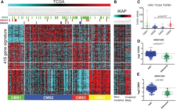
Human colorectal cancer (CRC) is a major cause of cancer mortality and frequently harbors activating mutations in the KRAS gene. To understand the role of oncogenic KRAS in CRC, we engineered a mouse model of metastatic CRC that harbors an inducible oncogenic Kras allele (Krasmut ) and conditional null alleles of Apc and Trp53 (iKAP). The iKAP model recapitulates tumor progression from adenoma through metastases. Whole-exome sequencing revealed that the Krasmut allele was heterogenous in primary tumors yet homogenous in metastases, a pattern consistent with activated Krasmut signaling being a driver of progression to metastasis. System-level and functional analyses revealed the TGF-β pathway as a key mediator of Krasmut -driven invasiveness. Genetic extinction of Krasmut resulted in specific elimination of the Krasmut subpopulation in primary and metastatic tumors, leading to apoptotic elimination of advanced invasive and metastatic disease. This faithful CRC model provides genetic evidence that Krasmut drives CRC invasion and maintenance of metastases.

Keywords: Apc; Kras; P53; colorectal cancer; invasion; metastasis.


Figures

Figure 1.
Figure 1.
iKAP GEMM of CRC. (A) GEMM design: Conditional alleles of Apc, Trp53, and tet-O-LSL-KrasG12D (Krasmut) were crossed with the tamoxifen-inducible VillinCreERT2, and 4-OHT tamoxifen was introduced into the adult colon via enema. (B) The model produces the full range of CRC progression, including macroscopic liver metastasis with Krasmut but not with Kraswt. (C) Median tumor-free survival is 6 mo and significantly longer without Krasmut. (D) Metastasis is dependent on Krasmut. (E) Endpoint tumors histologically staged by the TNM standard show that mutant Kras shifts the spectrum toward more invasive and malignant tumors. Bars: gross liver mets, 2 mm; histology, 500 µm; insets, 50 µm. (****) P < 0.0001 versus −C; (*) P < 0.05 versus −C; (NA) not applicable; (n.s.) not significant.
Figure 2.
Figure 2.
Kras clonal evolution. (A) Early stage iKAP tumors are heterogenous for Krasmut, but those cells predominate in late stage tumors and metastasis as visualized by native GFP fluorescence. (B) Immunohistochemistry (IHC) against GFP-tagged Krasmut cells showing Krasmut at the invasive front (red arrow) and increasing with tumor stage. (C) Quantification of GFP and pERK IHC signal across 251 tumors shows a dramatic selection for Krasmut cells at the invasive front of T1 adenocarcinomas and then throughout the bulk of T4 adenocarcinomas and metastases. Each tumor or invasive region was binned based on the percentage of GFP+ cells. (Ad) Adenoma; (AdC) adenocarcinoma; (Inv) invasive front; (Met) metastasis. Bars: 500 µm; insets, 50 µm.
Figure 3.
Figure 3.
Kras withdrawal in situ. (A) Mouse endoscopy showing iKAP tumor growth of tumors remaining on Dox (+Dox/+Kras) and after Dox withdrawal (−Dox/−Kras) within the same mouse over time. (B,C) Tumor growth was significantly reduced in −Dox/−Kras (n = 16) (B) as well as final tumor diameter (C) compared with “on Dox” (n = 48). (D,E) TNM staging of CRC tumors after random assignment to either remain +Dox/+Kras (D) or be taken off (−Dox/−Kras) (E) for 3 wk. The −Dox/−Kras tumors were exclusively adenomas, while the mice left on Dox had the full range of progression to metastasis. (F) H&E and IHC of iKAP tumors show GFP- and pERK-positive regions +Dox/+Kras. Upon Dox withdrawal for 2 d, GFP cells remained but no longer had activated pERK (arrows), and, after 2 wk off Dox, GFP-positive cells were mostly gone (‡), leaving large pockets of necrosis (†). (G) Biopsies were taken from iKAP tumors +Dox/+Kras, and then Dox was withdrawn for 2 wk. The previously biopsied tumors revealed that the GFP+/pERK+ population of cells was gone, and cCASP3-positive pockets of apoptotic cells were present (†). Bars: F, 500 µm; F inset, 50 µm; G, 200 µm; G inset, 50 µm. (**) P < 0.01; (*) P < 0.05.
Figure 4.
Figure 4.
Invasive iKAP tumor cells activate Tgfβ1 in a Krasmut -dependent manner. (A) Unsupervised hierarchical clustering of all mouse CRC samples showed similarity of expression by Krasmut status, KrasG12D transcript expression, and tumor stage. Three mice with T3/4 tumors show 0%–3% KRAS expression; we note that this is because the noninvasive regions were intentionally profiled. (B) Selected GSEA enrichment plots from analysis of the full 17,179-gene list from the in vivo data set. One-thousand-five-hundred-fifty pathways were significant at a false discovery rate (FDR) of <0.1. (C) Tgfβ1 expression in vivo. (D) Kras expression in vivo. (****) P < 0.0001; (n.s.) not significant.
Figure 5.
Figure 5.
TGF-β modulates invasion of iKAP cells. (A) Primary iKAP CRC cells cultured with Dox to induce Krasmut and with or without recombinant TGF-β and/or TGF-β inhibitor added to the medium. Crystal violet-stained invasion chambers showing recombinant TGF-β increases invasion when Krasmut is active. In Krasmut cells, invasion is increased further with additional TGF-β and decreased substantially by adding TGF-β inhibitor. (B) Invasion was quantified by counting migrated cells in three independent experiments. (C) Western blot on the same culture conditions as in A showing pSMAD2/3 as a readout of TGF-β pathway activation and pERK as a readout of RAS pathway activation. (D) IHC against GFP, TGFβ1, and pSMAD2 in invasive versus noninvasive tumors showing TGF-β activation in GFP+/Krasmut regions of tumors. Bars: 10×, 200 µm; 40×, 50 µm. (****) P < 0.0001.
Figure 6.
Figure 6.
iKAP tumors resemble human CRC subtype CMS4. (A) TCGA CRC expression data for the 416 genes in the CMS signature showing segmentation into the four distinct CMS subtypes across the bottom. Mutation status of KRAS, SMAD4, and SMAD2 in the TCGA samples is represented at the top. (B) iKAP mouse CRC expression data demonstrate that the invasive/metastatic iKAP tumors share the CMS4 signature, defined as mesenchymal with stromal invasion and high TGF-β signaling. (C) TGFβ1 expression in the human TCGA CRC data set, separated by CMS subtypes. (D) Quantification of TGFB1 expression comparing human adenocarcinomas (AdC) with adenomas in data set GSE41258. (E) Quantification of the CMS4 gene signature in the same data set.

References

    1. Anders S, Pyl PT, Huber W. 2015. HTSeq—a Python framework to work with high-throughput sequencing data. Bioinformatics 31: 166–169. - PMC - PubMed
    1. Belteki G, Haigh J, Kabacs N, Haigh K, Sison K, Costantini F, Whitsett J, Quaggin SE, Nagy A. 2005. Conditional and inducible transgene expression in mice through the combinatorial use of Cre-mediated recombination and tetracycline induction. Nucleic Acids Res 33: e51. - PMC - PubMed
    1. Chanrion M, Kuperstein I, Barriere C, El Marjou F, Cohen D, Vignjevic D, Stimmer L, Paul-Gilloteaux P, Bieche I, Tavares SD, et al. 2014. Concomitant Notch activation and p53 deletion trigger epithelial-to-mesenchymal transition and metastasis in mouse gut. Nat Commun 5: 5005. - PMC - PubMed
    1. Chin L, Tam A, Pomerantz J, Wong M, Holash J, Bardeesy N, Shen Q, O'Hagan R, Pantginis J, Zhou H, et al. 1999. Essential role for oncogenic Ras in tumour maintenance. Nature 400: 468–472. - PubMed
    1. David CJ, Huang YH, Chen M, Su J, Zou Y, Bardeesy N, Iacobuzio-Donahue CA, Massague J. 2016. TGF-β tumor suppression through a lethal EMT. Cell 164: 1015–1030. - PMC - PubMed

Substances