. 2018 Feb;23(2):304-315.
doi: 10.1038/mp.2017.37. Epub 2017 Mar 14.

The schizophrenia- and autism-associated gene, transcription factor 4 regulates the columnar distribution of layer 2/3 prefrontal pyramidal neurons in an activity-dependent manner

Affiliations

The schizophrenia- and autism-associated gene, transcription factor 4 regulates the columnar distribution of layer 2/3 prefrontal pyramidal neurons in an activity-dependent manner

S C Page et al. Mol Psychiatry. 2018 Feb.

Abstract

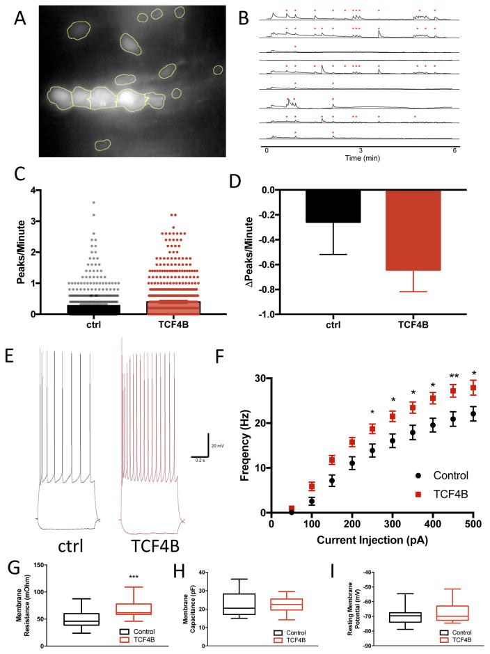
Disruption of the laminar and columnar organization of the brain is implicated in several psychiatric disorders. Here, we show in utero gain-of-function of the psychiatric risk gene transcription factor 4 (TCF4) severely disrupts the columnar organization of medial prefrontal cortex (mPFC) in a transcription- and activity-dependent manner. This morphological phenotype was rescued by co-expression of TCF4 plus calmodulin in a calcium-dependent manner and by dampening neuronal excitability through co-expression of an inwardly rectifying potassium channel (Kir2.1). For we believe the first time, we show that N-methyl-d-aspartate (NMDA) receptor-dependent Ca2+ transients are instructive to minicolumn organization because Crispr/Cas9-mediated mutation of NMDA receptors rescued TCF4-dependent morphological phenotypes. Furthermore, we demonstrate that the transcriptional regulation by the psychiatric risk gene TCF4 enhances NMDA receptor-dependent early network oscillations. Our novel findings indicate that TCF4-dependent transcription directs the proper formation of prefrontal cortical minicolumns by regulating the expression of genes involved in early spontaneous neuronal activity, and thus our results provides insights into potential pathophysiological mechanisms of TCF4-associated psychiatric disorders.


Conflict of interest statement

CONFLICT OF INTEREST

The authors declare no conflict of interest.

Figures

Figure 1
Figure 1. TCF4 gain-of-function results in abnormal columnar organization in the medial prefrontal cortex
TCF4 was expressed in the developing cortex via in utero electroporation. Brains of the resultant pups were fixed at p21 and imaged using confocal microscopy. TCF4 overexpression (A, A1) resulted in abnormal distribution of layer 2/3 pyramidal cells in medial prefrontal cortex (mPFC) when compared to GFP controls (A2). Knockdown of TCF4 expression by shTCF4 did not alter neuronal distribution (B, B2) compared to shCon (B2). This morphological phenotype was specific to the mPFC because no cellular distribution defects were observed when TCF4B was expressed in somatosensory cortex (SCx; C, C1, C2). The distribution of cells was quantified by binning pixel intensity perpendicular to the surface and calculating the coefficient of variance (CV) across all bins, therefore a high CV value indicates a more clustered distribution. D) Expression of TCF4B resulted in a significant increase in the CV when compared to GFP-only controls in the mPFC (GFP n=4 0.30±0.036 vs. TCF4B n=5 0.57±0.036; p<0.01). E. TCF4 loss-of-function by shRNA does not significantly alter pyramidal neuron distribution in mPFC (shCon n=4 0.29±0.035; shTCF4 n=5 0.34±0.012; p=0.18). F. In SCx, coefficient of variance is unchanged between GFP control and TCF4B (GFP n=5 0.295±0.014 vs. TCF4B n=4 0.27±0.017; p=0.18). Scale bar 100μm, inset 50 μm.
Figure 2
Figure 2. TCF4-dependent regulation of cellular distribution requires transcription
A) Diagram of three different TCF4 isoforms depicting the known protein motifs which include two activation domains (AD1 and AD2), a nuclear localization sequence (NLS) and the DNA-binding basic Helix-Loop-Helix domain (bHLH). B) TCF4B produces pyramidal cell aggregation in the mPFC, as does the short isoform TCF4A that lacks a NLS. A disease-causing point mutant, R582P, does not alter pyramidal cell distribution. Co-expression of TCF4B + R582P rescues TCF4B-dependent formation of abnormal minicolumn structures. Expression of TCF4B that lacks an AD2 (ΔAD2) does not disrupt pyramidal cell distribution. Abnormal column formation is not observed when the TCF4H isoform is overexpressed. Scale bar 50μm. C) Group analysis indicates that R582P (n=4, 0.33±0.036, p=0.0007), TCF4B + R582P (n=5, 0.38±0.025, p=0.021), ΔAD2 (n=6, 0.26±0.017; p<0.001), and TCF4H (n=8, 0.31±0.030, p<0.001) cause a significant reduction in CV compared to TCF4B (n=6, 0.57±0.061) while expression of TCF4A (n=9, 0.45±0.039, p>0.05; ANOVA p<0.0001) does not significantly reduce CV value when compared to TCF4B. *P<0.05, **P<0.01, ***P<0.001, ****P<0.0001
Figure 3
Figure 3. Abnormal column formation is rescued by co-expression of calmodulin (CaM) and this rescue requires calcium binding to CaM
A) TCF4B was expressed alone, co-expressed with CaM or with a mutant calmodulin that is unable to bind Ca2+ (CaM1,2,3,4); CaM (n=6) or CaM1,2,3,4 (n=4) were also expressed alone as controls. Scale bar 50μm. B) Cortical column formation was significantly reduced in the TCF4B + CaM condition (TCF4B n=6, 0.51±0.049 vs. TCF4B + CaM n=5, 0.31±0.012, p=0.028), the CaM condition (n=6, 0.18±0.019, p=0.001), or the CaM1,2,3,4 condition (n=4, 0.25±0.057, p=0.004) but not in the TCF4B + CaM1,2,3,4 condition (TCF4B + CaM1,2,3,4 n=5, 0.56±0.071; p>0.05; ANOVA p<0.0001). C) Co-immunoprecipitation experiments demonstrate that myc-TCF4B can immunoprecipitate both CaM (left) and CaM1,2,3,4 (right). D) Co-immunoprecipitation is observed regardless of the amount of Ca2+ present during the immunoprecipitation reaction.
Figure 4
Figure 4. TCF4-induced abnormal column formation is activity-and NMDA receptor-dependent
A) An inwardly rectifying potassium channel (Kir2.1) rescues TCF4B-dependent disruption of pyramidal cell distribution. A mutant Kir2.1 channel (Kir2.1M) that is expressed on the membrane but does not pass current was ineffective at rescue. Expression of either channel alone has no effect on cellular distribution. Scale bar 50μm. B) Group analysis showing that co-expression of TCF4B + Kir2.1 prevents abnormal column formation (TCF4B n=6, 0.46±0.065 vs. TCF4B + Kir2.1 n=5, 0.23±0.022; p=0.012; ANOVA p=0.007). TCF4B + Kir2.1M did not prevent column formation (TCF4B + Kir2.1M n=6, 0.42±0.051, p>0.05), while either Kir2.1 (n=4, 0.24±0.020; p=0.032) or Kir2.1M (n=5, 0.28±0.046, p=0.07) alone had no effect on cellular distribution. (** P<0.01; * P<0.05; # P<0.10). C) TCF4B-dependent cellular aggregation was observed when TCF4B was co-expressed with a Crispr/Cas9 vector lacking a guide RNA that, when expressed alone, did not result in cellular aggregation. (TCF4B + crEmpty, n=5 0.73±0.11 vs. crEmpty, n=5, 0.27±0.036; p<0.001; ANOVA p<0.0001). Co-expression TCF4B with a Crispr/Cas9 construct that targets Grin1 rescues cellular aggregation, while co-expression of a Crispr/Cas9 that targets the AMPA receptor subunit Gria2 failed at rescue of cellular aggregation. Scale bar 50μm. D) Cellular distribution analysis indicates TCF4B + crGrin1 significantly rescued pyramidal cell aggregation compared to TCF4B + crEmpty (TCF4B + crGrin1 n=5, 0.36±0.024, p<0.0001), and TCF4B + crGria2 did not rescue TCF4B-dependent cellular aggregation (TCF4B + crGria2 n=4, 0.55±0.009, p>0.05) Expression of crGrin1 (n=5, 0.29±0.032, p<0.001) or crGria2 (n=5, 0.33±0.033, p=0.002) alone has no effect on pyramidal cell distribution (** P<0.01; ****p<0.0001).
Figure 5
Figure 5. TCF4B gain of function significantly enhances spontaneous neuronal activity
A) Example image of an abnormal minicolumn structure in pyramidal neurons expressing TCF4B + mCherry + GCaMP6s. A custom R script was written to identify individual cells (ROI) using the mCherry channel. B) Example traces of spontaneous Ca2+ events that were detected (*). Pixel intensity was normalized to background for each image, and peaks were called using a custom R script. C) Total activity was calculated by number of peaks per cell per minute, and was significantly increased in the TCF4B condition compared to control (TCF4B n=743 cells, 0.3950±0.0202 vs. Ctrl n=450, 0.3020±0.0232; t-test; P<0.001). D) Ca2+ transients were inhibited by bath application of the NMDA receptor antagonist DL-AP5 (100μM). A Poisson mix effect model was used to discern the difference in activity by DL-AP5 with each transfection condition. Addition of DL-AP5 inhibited activity both transfection conditions (TCF4B n=201, −0.2140±0.0294; ctrl n=91, −0.0857±0.0248; p<4.06e-06). TCF4B transfect cells were more affected by AP5 compared to ctrl cells (post-hoc t-test; P<0.00655). E). Sample current-clamp traces recorded from GFP- or TCF4B-expressing neurons showing increased action potential output in response to current injection (−50 pA, +400 pA). F) Summary data shows that the frequency of action potentials increases in response to increasing current injection. Post hoc analysis indicated that TCF4B cells (n=27) produced significantly more spikes than GFP control cells (n=19) between 300 and 500 pA of current injection. G) TCF4B cells show in an increase in membrane resistance compared to control (control n=19, 49.94±4.223: TCF4B n=27, 68.67±2.829; p=0.004). H) Membrane capacitance is unchanged between TCF4B and control cells (control n=19, 22.96±1.565: TCF4B n=27, 21.89±0.877; p=0.525). I) Resting membrane potential is unchanged between TCF4B and control cells (control n=19, −69.94±1.387: TCF4B n=27, −67.74±1.178; p=0.238).

References

    1. Casanova MF, Buxhoeveden DP, Switala AE, Roy E. Minicolumnar pathology in autism. Neurology. 2002;58:428–432. - PubMed
    1. Casanova MF, Buxhoeveden DP, Cohen M, Switala AE, Roy EL. Minicolumnar pathology in dyslexia. Ann Neurol. 2002;52:108–110. - PubMed
    1. Casanova MF, de Zeeuw L, Switala A, Kreczmanski P, Korr H, Ulfig N, et al. Mean cell spacing abnormalities in the neocortex of patients with schizophrenia. Psychiatry Research. 2005;133:1–12. - PubMed
    1. Gleeson JG. Classical lissencephaly and double cortex (subcortical band heterotopia): LIS1 and doublecortin. Curr Opin Neurol. 2000;13:121–125. - PubMed
    1. Belmonte MK. Autism and Abnormal Development of Brain Connectivity. Journal of Neuroscience. 2004;24:9228–9231. - PMC - PubMed

Publication types

Substances