. 2017 Feb 24:7:27-37.
doi: 10.1016/j.ynstr.2017.02.004. eCollection 2017 Dec.

Cav1.2 channels mediate persistent chronic stress-induced behavioral deficits that are associated with prefrontal cortex activation of the p25/Cdk5-glucocorticoid receptor pathway

Affiliations

Cav1.2 channels mediate persistent chronic stress-induced behavioral deficits that are associated with prefrontal cortex activation of the p25/Cdk5-glucocorticoid receptor pathway

Charlotte C Bavley et al. Neurobiol Stress. .

Abstract

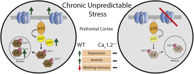
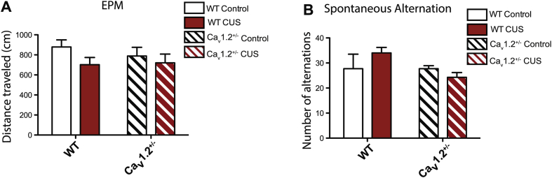
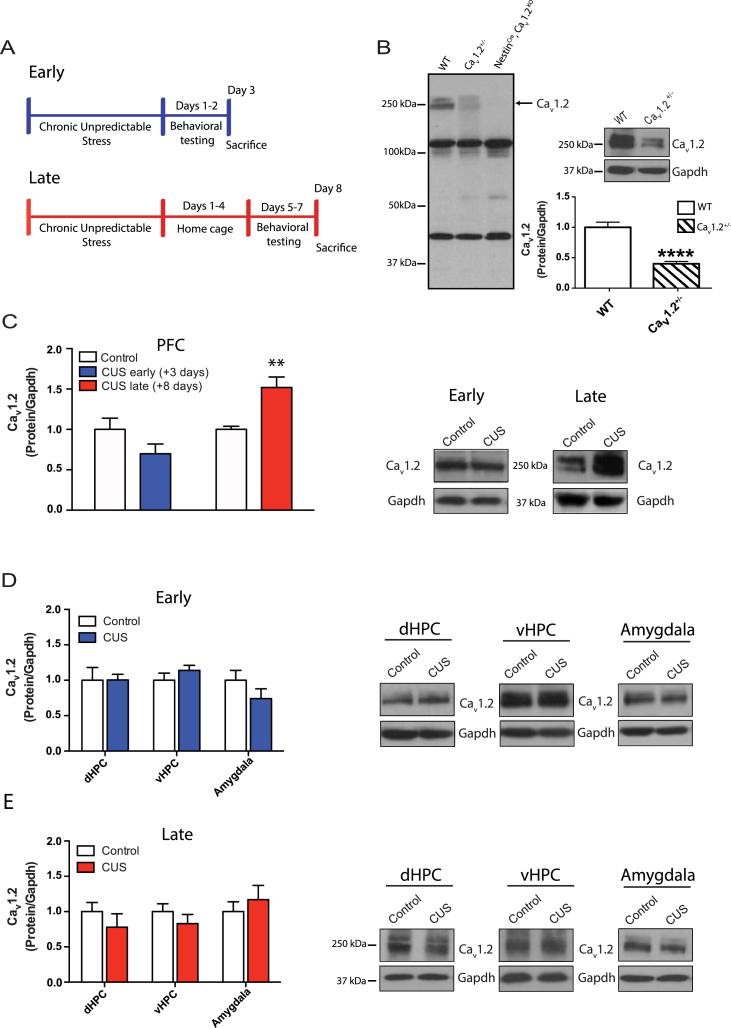
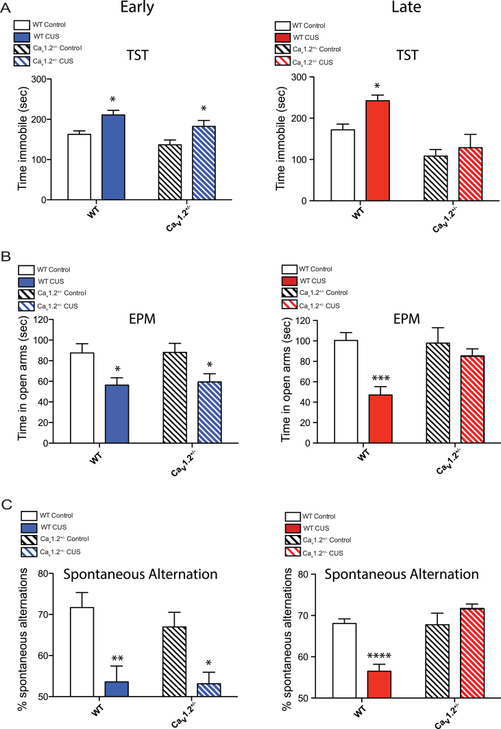
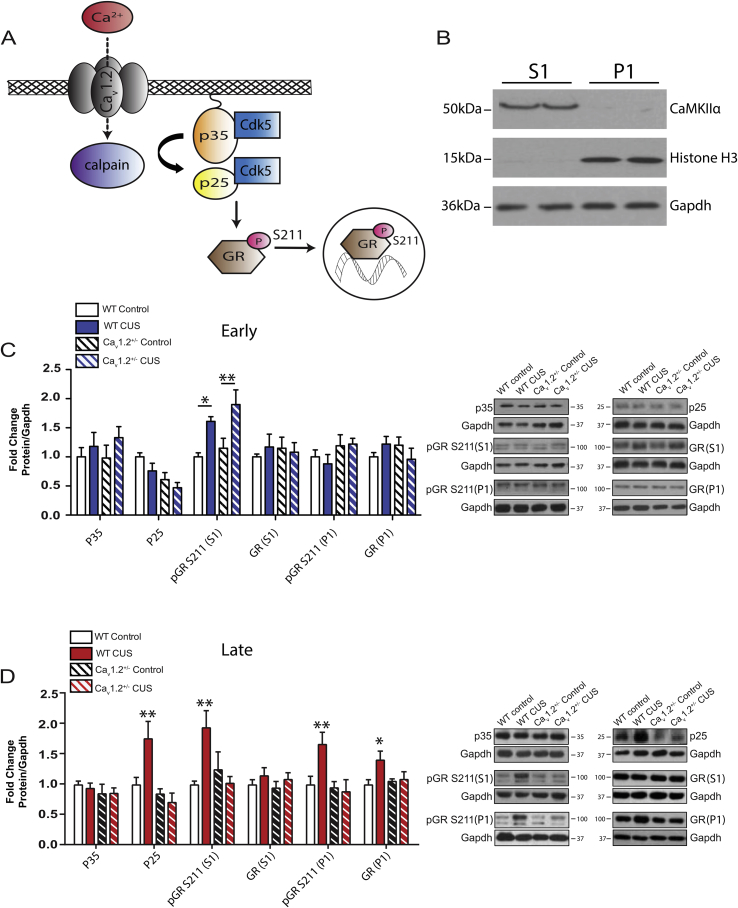
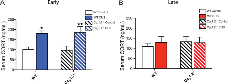
Chronic stress is known to precipitate and exacerbate neuropsychiatric symptoms, and exposure to stress is particularly pathological in individuals with certain genetic predispositions. Recent genome wide association studies have identified single nucleotide polymorphisms (SNPs) in the gene CACNA1C, which codes for the Cav1.2 subunit of the L-type calcium channel (LTCC), as a common risk variant for multiple neuropsychiatric conditions. Cav1.2 channels mediate experience-dependent changes in gene expression and long-term synaptic plasticity through activation of downstream calcium signaling pathways. Previous studies have found an association between stress and altered Cav1.2 expression in the brain, however the contribution of Cav1.2 channels to chronic stress-induced behaviors, and the precise Cav1.2 signaling mechanisms activated are currently unknown. Here we report that chronic stress leads to a delayed increase in Cav1.2 expression selectively within the prefrontal cortex (PFC), but not in other stress-sensitive brain regions such as the hippocampus or amygdala. Further, we demonstrate that while Cav1.2 heterozygous (Cav1.2+/-) mice show chronic stress-induced depressive-like behavior, anxiety-like behavior, and deficits in working memory 1-2 days following stress, they are resilient to the effects of chronic stress when tested 5-7 days later. Lastly, molecular studies find a delayed upregulation of the p25/Cdk5-glucocorticoid receptor (GR) pathway in the PFC when examined 8 days post-stress that is absent in Cav1.2+/- mice. Our findings reveal a novel Cav1.2-mediated molecular mechanism associated with the persistent behavioral effects of chronic stress and provide new insight into potential Cav1.2 channel mechanisms that may contribute to CACNA1C-linked neuropsychiatric phenotypes.


Figures

Image 1
Graphical abstract
Bavley et al._Supp Figure 1-05
Behavioral analyses of Cav1.2 heterozygous (Cav1.2+/−) and wildtype littermates (WT) following exposure to chronic unpredictable stress (CUS). A) No effect of genotype or CUS was observed on distance traveled in the elevated plus maze (EPM) (genotype: F(1,45) = 0.20, p = 0.66; CUS: F(1,45) = 2.36, p = 0.13; WT control, n = 12; WT CUS, n = 14; Cav1.2+/− control, n = 10; Cav1.2+/− CUS, n = 13). B) No effect of genotype or CUS was observed on total number of alternations in the spontaneous alternation task (genotype: F(1,24) = 2.08, p = 0.16; CUS: F(1,24) = 0.19, p = 0.67; WT control, n = 7; WT CUS, n = 8; Cav1.2+/− control, n = 7; Cav1.2+/− CUS, n = 7). Data are represented as mean ± SEM.
Bavley et al._Supp Figure 2-06
Western blot analysis of the p25/ Cdk5-GR pathway in the dHPC, vHPC, and amygdala following exposure to chronic unpredictable stress (CUS) in WT and Cav1.2+/− mice. A) CUS increased phosphorylation of the glucocorticoid receptor (GR) at S211 in the dHPC when examined at the early (3 days post-stress) time point (t(7) = 4.44, **p = 0.003) but had no effect on p25 or GR expression (control, n = 7; CUS, n = 7). In the vHPC and amygdala, CUS had no effect on protein levels of p25, pGR S211 or GR when examined at the early time point (control, n = 7; CUS, n = 7). B) CUS had no effect on protein levels of p25, pGR S211 or GR in the dHPC, vHPC or amygdala when examined at late (8 days post-stress) time point (control, n = 6; CUS, n = 5–7). Data are represented as mean ± SEM.
Fig. 1
Fig. 1
Western blot analysis of Cav1.2 expression in the prefrontal cortex following chronic unpredictable stress. A) Schematics of the chronic unpredictable stress paradigms, indicating the early and late time points chosen. B) Left, Validation of the Cav1.2 antibody using a full brain knockout (NestinCre, Cav1.2KO) and Cav1.2+/− mice indicates that the two bands surrounding 250 kDa are specific Cav1.2 variants. Right, When quantified, these two bands are significantly reduced in Cav1.2+/− PFC (****p < 0.0001; WT, n = 5; Cav1.2+/−, n = 6). C) Chronic unpredictable stress (CUS) did not affect Cav1.2 protein expression in the PFC when examined at the early (3 days post-stress) time point, but increased Cav1.2 protein expression in the PFC when examined at the late (8 days post-stress) time point (**p < 0.01; control, n = 6; CUS, n = 7). Western blot images contain representative samples of each group taken from the same blot. D) CUS does not impact Cav1.2 protein levels in the dHPC (control, n = 7; CUS, n = 6), vHPC (control, n = 7; CUS, n = 7), or amygdala (control, n = 7; CUS, n = 7) when examined at the early time point. E) CUS does not impact Cav1.2 protein levels in the dHPC (control, n = 6; CUS, n = 5), vHPC (control, n = 6; CUS, n = 6) or amygdala (control, n = 6; CUS, n = 5) when examined at the late time point. Data are represented as mean ± SEM.
Fig. 2
Fig. 2
Behavior of Cav1.2 heterozygous (Cav1.2+/−) and wildtype littermates (WT) following exposure to chronic unpredictable stress (CUS). A) Left, CUS increased depressive-like behavior in the tail suspension test (TST) in both WT and Cav1.2+/− mice when tested at the early (1–2 days post-stress) time point (*p < 0.05, WT control vs CUS, bonferroni post-hoc; *p < 0.05, Cav1.2+/− control vs CUS, bonferroni post-hoc; WT control, n = 6; WT CUS, n = 7; Cav1.2+/− control, n = 8; Cav1.2+/− CUS, n = 7). Right, when tested at the late (5–7 days post-stress) time piont, CUS increased depressive-like behavior in WT mice but not in Cav1.2+/− mice (*p < 0.05, WT control vs CUS, bonferroni post-hoc; p > 0.05, Cav1.2+/− control vs CUS, bonferroni post-hoc; WT control, n = 5; WT CUS, n = 8; Cav1.2+/− control, n = 5; Cav1.2+/− CUS, n = 5). B) Left, when tested at the early time point, CUS increased anxiety-like behavior in the elevated plus maze (EPM) in both WT and Cav1.2+/− mice (*p < 0.05, WT control vs CUS, bonferroni post-hoc; *p < 0.05, Cav1.2+/− control vs CUS, bonferroni post-hoc; WT control, n = 7; WT CUS, n = 7; Cav1.2+/− control, n = 8; Cav1.2+/− CUS, n = 7). Right, when tested at the late time point, CUS increased anxiety-like behavior in WT but not in Cav1.2+/− mice (***p < 0.001, WT control vs CUS, bonferroni post-hoc; p = 0.60, Cav1.2+/− control vs CUS, bonferroni post-hoc; WT control, n = 12; WT CUS, n = 14; Cav1.2+/− control, n = 10; Cav1.2+/− CUS, n = 13). C) Left, CUS induced working memory deficits in the spontaneous alternation task in WT and Cav1.2+/− mice when tested at the early time point (**p < 0.01, WT control vs CUS, bonferroni post-hoc; *p < 0.05, Cav1.2+/− control vs CUS, bonferroni post-hoc; WT control, n = 7; WT CUS, n = 7; Cav1.2+/− control, n = 8; Cav1.2+/− CUS, n = 7). Right, when tested at the late time point, however, CUS decreased spontaneous alternations in WT but not in Cav1.2+/− mice (****p < 0.0001 WT control vs CUS, bonferroni post-hoc; p = 0.22, Cav1.2+/− control vs CUS, bonferroni post-hoc; WT control, n = 12; WT CUS, n = 14; Cav1.2+/− control, n = 10; Cav1.2+/− CUS, n = 13). Data are represented as mean ± SEM.
Fig. 3
Fig. 3
Western blot analysis of the p25/Cdk5-GR pathway in the PFC following exposure to chronic unpredictable stress (CUS) in WT and Cav1.2+/− mice. A) Working model of Cav1.2 activation of the p25/Cdk5-GR pathway. B) Representative western blots with S1 (cytoplasmic and membrane) and P1 (nuclear) fractions demonstrating purity of fractions using the S1 and P1 fraction-specific proteins CaMKIIα and Histone H3, respectively. C) In the PFC, CUS increased expression of phosphorylated glucocorticoid receptors at S211 (pGR S211) in the S1 fraction in both WT and Cav1.2+/− mice (*p < 0.05, WT control vs CUS, bonferroni post-hoc; **p < 0.01, Cav1.2+/− control vs CUS, bonferroni post-hoc) when examined at the early (3 days post-stress) time point. At this time point, CUS had no effect on expression of p35, p25, GR in the S1 fraction, pGR S211 in the P1 fraction or GR in the P1 fraction (WT control, n = 5–7; WT CUS, n = 7; Cav1.2+/− control, n = 7; Cav1.2+/− CUS, n = 6–7). D) When examined at the late (8 days post-stress) time point, in the PFC of WT but not Cav1.2+/− mice, CUS increased expression of p25 (WT control vs CUS, **p < 0.01, bonferroni post-hoc; Cav1.2+/− control vs CUS, p > 0.99, bonferroni post-hoc; WT control, n = 11; WT CUS, n = 10; Cav1.2+/− control, n = 9; Cav1.2+/− CUS, n = 11), phosphorylation of glucocorticoid receptors at S211 (pGR S211) in both the S1 fraction (WT control vs CUS, **p < 0.01, bonferroni post-hoc; Cav1.2+/− control vs CUS, p = 0.70, bonferroni post-hoc; WT control, n = 6; WT CUS, n = 7; Cav1.2+/− control, n = 7; Cav1.2+/− CUS, n = 7) and in the P1 fraction (**p < 0.01 WT control vs CUS, bonferroni post-hoc; p = 0.96 Cav1.2+/− control vs CUS, bonferroni post-hoc; WT control, n = 6; WT CUS, n = 8; Cav1.2+/− control, n = 6; Cav1.2+/− CUS, n = 5), as well as P1 levels of GR (*p < 0.05 WT control vs CUS, bonferroni post-hoc; p = 0.85 Cav1.2+/− control vs CUS, bonferroni post-hoc; WT control, n = 6; WT CUS, n = 8; Cav1.2+/− control, n = 6; Cav1.2+/− CUS, n = 6). CUS had no effect on S1 expression of p35 or GR in either WT or Cav1.2+/− mice (WT control, n = 6–8; WT CUS, n = 7; Cav1.2+/− control, n = 6–7; Cav1.2+/− CUS, n = 7). Western blot images contain representative samples of each group taken from the same blot. Data are represented as mean ± SEM.
Fig. 4
Fig. 4
Analysis of serum corticosterone concentration following acute and chronic stress. A) Chronic unpredictable stress (CUS) increased serum corticosterone (CORT) levels in both WT and Cav1.2+/− mice when examined at the early (3 days post-stress) time point (*p < 0.05, WT control vs CUS, bonferroni post-hoc; **p < 0.01, Cav1.2+/− control vs CUS, bonferroni post-hoc; WT control, n = 7; WT CUS, n = 7; Cav1.2+/− control, n = 8; Cav1.2+/− CUS, n-6). B) When examined at the late (8 days post-stress) time point, (CUS had no effect on serum CORT levels in either WT or Cav1.2+/− mice (WT control, n = 9; WT CUS, n = 14; Cav1.2+/− control, n = 11; Cav1.2+/− CUS, n = 13). Data are represented as mean ± SEM.

References

    1. Adhikari A., Topiwala M.A., Gordon J.A. Synchronized activity between the ventral hippocampus and the medial prefrontal cortex during anxiety. Neuron. 2010;65:257–269. - PMC - PubMed
    1. Adhikari A., Lerner T.N., Finkelstein J., Pak S., Jennings J.H., Davidson T.J., Ferenczi E., Gunaydin L.A., Mirzabekov J.J., Ye L., Kim S.Y., Lei A., Deisseroth K. Basomedial amygdala mediates top-down control of anxiety and fear. Nature. 2015;527:179–185. - PMC - PubMed
    1. Adzic M., Djordjevic J., Djordjevic A., Niciforovic A., Demonacos C., Radojcic M., Krstic-Demonacos M. Acute or chronic stress induce cell compartment-specific phosphorylation of glucocorticoid receptor and alter its transcriptional activity in Wistar rat brain. J. Endocrinol. 2009;202:87–97. - PMC - PubMed
    1. Ament S.A., Szelinger S., Glusman G., Ashworth J., Hou L., Akula N., Shekhtman T., Badner J.A., Brunkow M.E., Mauldin D.E., Stittrich A.B., Rouleau K., Detera-Wadleigh S.D., Nurnberger J.I., Jr., Edenberg H.J., Gershon E.S., Schork N., Bipolar Genome Study, Price ND. Gelinas R., Hood L., Craig D., McMahon F.J., Kelsoe J.R., Roach J.C. Rare variants in neuronal excitability genes influence risk for bipolar disorder. Proc. Natl. Acad. Sci. 2015;112:3576–3581. (U.S.A) - PMC - PubMed
    1. Bader P.L., Faizi M., Kim L.H., Owen S.F., Tadross M.R., Alfa R.W., Bett G.C., Tsien R.W., Rasmusson R.L., Shamloo M. Mouse model of Timothy syndrome recapitulates triad of autistic traits. Proc. Natl. Acad. Sci. 2011;108:15432–15437. (U.S.A.) - PMC - PubMed

LinkOut - more resources