. 2017 Mar 9;2(5):e89530.
doi: 10.1172/jci.insight.89530.

ALS patients' regulatory T lymphocytes are dysfunctional, and correlate with disease progression rate and severity

Affiliations

ALS patients' regulatory T lymphocytes are dysfunctional, and correlate with disease progression rate and severity

David R Beers et al. JCI Insight. .

Abstract

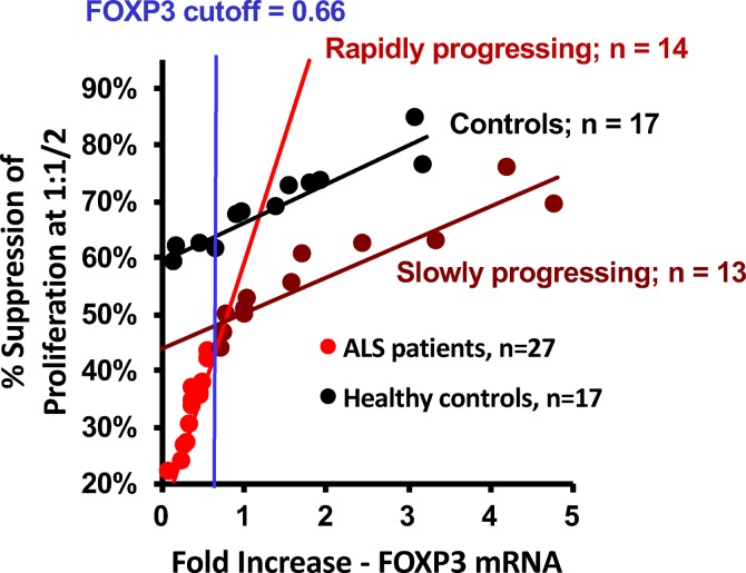
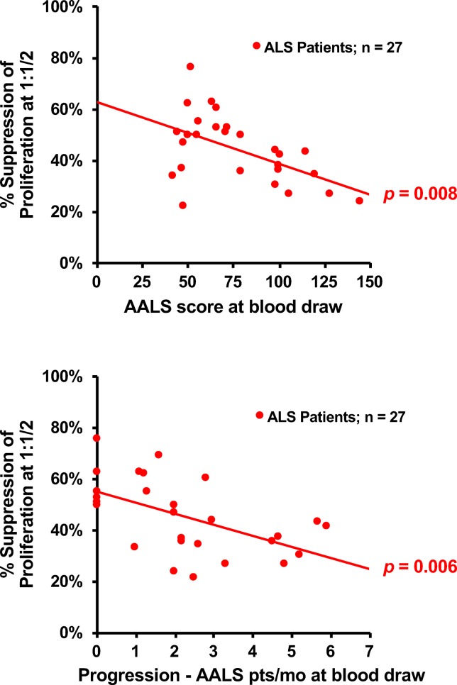
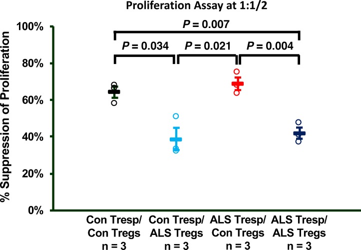
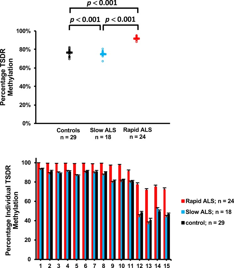
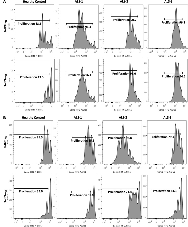
Neuroinflammation is a pathological hallmark of ALS in both transgenic rodent models and patients, and is characterized by proinflammatory T lymphocytes and activated macrophages/microglia. In ALS mouse models, decreased regulatory T lymphocytes (Tregs) exacerbate the neuroinflammatory process, leading to accelerated motoneuron death and shortened survival; passive transfer of Tregs suppresses the neuroinflammation and prolongs survival. Treg numbers and FOXP3 expression are also decreased in rapidly progressing ALS patients. A key question is whether the marked neuroinflammation in ALS can be attributed to the impaired suppressive function of ALS Tregs in addition to their decreased numbers. To address this question, T lymphocyte proliferation assays were performed. Compared with control Tregs, ALS Tregs were less effective in suppressing responder T lymphocyte proliferation. Although both slowly and rapidly progressing ALS patients had dysfunctional Tregs, the greater the clinically assessed disease burden or the more rapidly progressing the patient, the greater the Treg dysfunction. Epigenetically, the percentage methylation of the Treg-specific demethylated region was greater in ALS Tregs. After in vitro expansion, ALS Tregs regained suppressive abilities to the levels of control Tregs, suggesting that autologous passive transfer of expanded Tregs might offer a novel cellular therapy to slow disease progression.


Conflict of interest statement

Conflict of interest: The authors have declared that no conflict of interest exists.

Figures

Figure 1
Figure 1. Tregs from ALS patients are dysfunctional.
(A) Scatter plot of the Treg suppression assay results using Tregs from individual controls and ALS patients. Tregs from patients with ALS are not as functionally suppressive as Tregs from control individuals. Significance determined by Student’s t test. (B) Tregs from patients with slowly progressing ALS and the control individuals are more suppressive than patients with rapidly progressing ALS across all responder T lymphocyte/Treg suppression assay ratios (CD4+CD25/CD4+CD25hi). C, control; S, slowly progressing ALS patients; R, rapidly progressing ALS patients.
Figure 2
Figure 2. The correlation of Treg suppression versus FOXP3 mRNA expression levels.
The percentage Treg suppression of responder T lymphocyte (Tresp) proliferation at the 1:1/2 Tresp/Treg ratio was very strongly correlated with FOXP3 mRNA expression.
Figure 3
Figure 3. Treg suppressive deficiency correlates with disease burden and rate of disease progression.
(A) There is a correlation between burden of disease (Appel ALS [AALS] points) at blood draw and the level of Treg suppression of responder T lymphocyte (Tresp) proliferation at the 1:1/2 Tresp/Treg ratio. (B) There is a correlation between the rates of disease progression (AALS points/month) at blood draw and Treg suppression of Tresp proliferation. P values obtained by Pearson correlation.
Figure 4
Figure 4. Dysfunction of ALS responder T lymphocytes (Tresps) versus control Tresps.
Mixing studies demonstrated that when assayed at a 1:1/2 Tresp/Treg ratio, Tregs from patients with ALS could not suppress the proliferative response of target Tresps from either ALS patients or the controls (Con). Significance determined by Student’s t test.
Figure 5
Figure 5. The percentage methylation of the Treg-specific demethylated region (TSDR) between ALS patients’ and controls’ Tregs.
(A) The percentage TSDR methylation combining all 15 CpG islands among the 3 groups. ALS patients exhibited more methylation in the TSDR of Tregs compared with the TSDR of slowly progressing ALS patients and the control group. Significance determined by Student’s t test. (B) The percentage TSDR methylation of each individual CpG island among the 3 groups. The TSDR methylation was especially enhanced in islands 12 through 15 in the rapidly progressing ALS patients compared with slowly progressing ALS patient and the control group. There were no differences across all 15 CpG island between slowly progressing ALS patients and the control group.
Figure 6
Figure 6. In vitro expansion induces a recovery of ALS Tregs’ suppressive capacities.
(A) Prior to expansion, ALS patients’ Tregs were again found to be less capable of suppressing their corresponding autologous CD4+CD25 responder T lymphocytes (Tresps) at a 1:1 Tresp/Treg ratio than the control group’s Tregs were of suppressing their corresponding autologous Tresps. (B) After expansion, the ALS patients’ Tregs acquired the ability to suppress their corresponding autologous Tresps to nearly same degree as the control group’s Tregs at a 1:1 Tresp/Treg ratio.

References

    1. Zhao W, Beers DR, Appel SH. Immune-mediated mechanisms in the pathoprogression of amyotrophic lateral sclerosis. J Neuroimmune Pharmacol. 2013;8(4):888–899. doi: 10.1007/s11481-013-9489-x. - DOI - PMC - PubMed
    1. Appel SH, Beers DR, Henkel JS. T cell-microglial dialogue in Parkinson’s disease and amyotrophic lateral sclerosis: are we listening? Trends Immunol. 2010;31(1):7–17. doi: 10.1016/j.it.2009.09.003. - DOI - PMC - PubMed
    1. Henkel JS, et al. Presence of dendritic cells, MCP-1, and activated microglia/macrophages in amyotrophic lateral sclerosis spinal cord tissue. Ann Neurol. 2004;55(2):221–235. doi: 10.1002/ana.10805. - DOI - PubMed
    1. Henkel JS, Beers DR, Siklós L, Appel SH. The chemokine MCP-1 and the dendritic and myeloid cells it attracts are increased in the mSOD1 mouse model of ALS. Mol Cell Neurosci. 2006;31(3):427–437. doi: 10.1016/j.mcn.2005.10.016. - DOI - PubMed
    1. Henkel JS, Beers DR, Zhao W, Appel SH. Microglia in ALS: the good, the bad, and the resting. J Neuroimmune Pharmacol. 2009;4(4):389–398. doi: 10.1007/s11481-009-9171-5. - DOI - PubMed

Publication types

MeSH terms