

Primary and metastatic ovarian cancer: Characterization by 3.0T diffusion-weighted MRI
- PMID: 28289938
- PMCID: PMC5544807
- DOI: 10.1007/s00330-017-4786-z
Primary and metastatic ovarian cancer: Characterization by 3.0T diffusion-weighted MRI
Abstract
Objectives: We aimed to investigate whether apparent diffusion coefficients (ADCs) measured by 3.0T diffusion-weighted magnetic resonance imaging (DWI) associate with histological aggressiveness of ovarian cancer (OC) or predict the clinical outcome. This prospective study enrolled 40 patients with primary OC, treated 2011-2014.
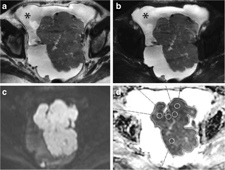
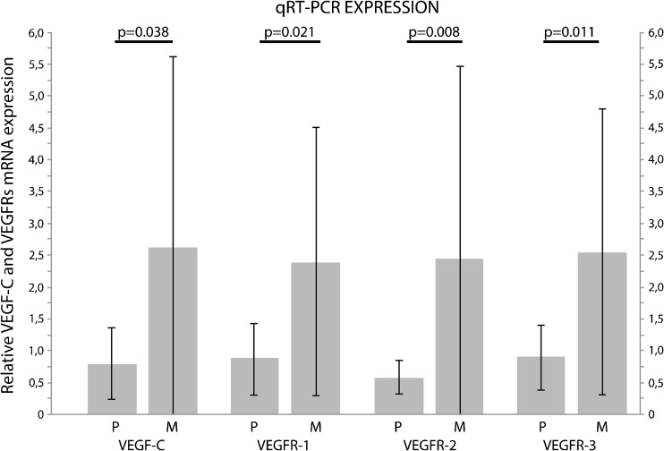
Methods: DWI was performed prior to surgery. Two observers used whole lesion single plane region of interest (WLsp-ROI) and five small ROIs (S-ROI) to analyze ADCs. Samples from tumours and metastases were collected during surgery. Immunohistochemistry and quantitative reverse transcription polymerase chain reaction (qRT-PCR) were used to measure the expression of vascular endothelial growth factor (VEGF) and its receptors.
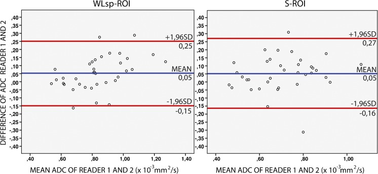
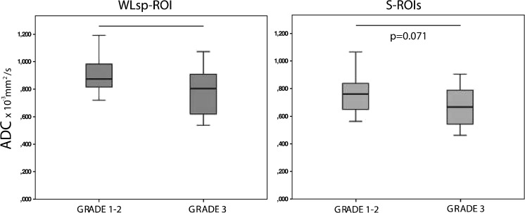
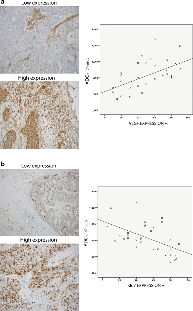
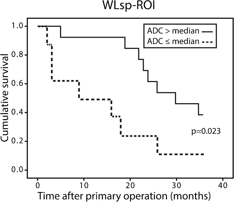
Results: The interobserver reliability of ADC measurements was excellent for primary tumours ICC 0.912 (WLsp-ROI). Low ADCs significantly associated with poorly differentiated OC (WLsp-ROI P = 0.035). In primary tumours, lower ADCs significantly associated with high Ki-67 (P = 0.001) and low VEGF (P = 0.001) expression. In metastases, lower ADCs (WLsp-ROI) significantly correlated with low VEGF receptors mRNA levels. ADCs had predictive value; 3-year overall survival was poorer in patients with lower ADCs (WLsp-ROI P = 0.023, S-ROI P = 0.038).
Conclusion: Reduced ADCs are associated with histological severity and worse outcome in OC. ADCs measured with WLsp-ROI may serve as a prognostic biomarker of OC.
Key points: • Reduced ADCs correlate with prognostic markers: poor differentiation and high Ki-67 expression • ADCs also significantly correlated with VEGF protein expression in primary tumours • Lower ADC values are associated with poorer survival in ovarian cancer • Whole lesion single plane-ROI ADCs may be used as a prognostic biomarker in OC.
Keywords: Cell proliferation; Diffusion magnetic resonance imaging; Neoplasm metastasis; Neovascularization pathologic; Ovarian neoplasms.
Conflict of interest statement
Guarantor
The scientific guarantor of this publication is Maarit Anttila.
Conflict of interest
The authors of this manuscript declare no relationships with any companies, whose products or services may be related to the subject matter of the article.
Funding
This study has received funding by the Finnish Medical Foundation, Kuopio University Hospital (VTR grant), Kuopio University Hospital Research Foundation and University of Eastern Finland.
Statistics and biometry
Statistician Tuomas Selander kindly provided statistical advice for this manuscript.
Ethical approval
Institutional Review Board approval was obtained.
Informed consent
Written informed consent was obtained from all subjects (patients) in this study.
Methodology
• prospective
• observational/experimental
• performed at one institution
Figures
References
MeSH terms
Substances
LinkOut - more resources
Full Text Sources
Other Literature Sources
Medical
