. 2017 Mar 14:7:44313.
doi: 10.1038/srep44313.

Structural Basis of Eco1-Mediated Cohesin Acetylation

Affiliations

Structural Basis of Eco1-Mediated Cohesin Acetylation

William C H Chao et al. Sci Rep. .

Erratum in

Abstract

Sister-chromatid cohesion is established by Eco1-mediated acetylation on two conserved tandem lysines in the cohesin Smc3 subunit. However, the molecular basis of Eco1 substrate recognition and acetylation in cohesion is not fully understood. Here, we discover and rationalize the substrate specificity of Eco1 using mass spectrometry coupled with in-vitro acetylation assays and crystallography. Our structures of the X. laevis Eco2 (xEco2) bound to its primary and secondary Smc3 substrates demonstrate the plasticity of the substrate-binding site, which confers substrate specificity by concerted conformational changes of the central β hairpin and the C-terminal extension.


Conflict of interest statement

The authors declare no competing financial interests.

Figures
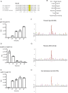
Figure 1
Figure 1. Smc3 K113 is the primary target for Eco1 and Eco1 substrate motif consensuses.
(A) Sequence alignment of Smc3 β hairpin region with the conserved tandem lysines highlighted. (B) Schematic of in-vitro Eco1-mediated acetylation time-course assay coupled to MS analysis. (C) In-vitro Eco1-mediated acetylation assay showing increased level of tandem lysine (K112 & K113) acetylation peptide over time. Area is in arbitrary unit. (D) In-vitro Eco1-mediated acetylation assay showing decreased level of unacetylated (dimethylated on both K112 and K113) peptide over time. (E) In-vitro Eco1-mediated acetylation assay showing rapid increase of sole- K113 acetylation peptide from 0 min to 10 min and approaching completion over time. This suggests that a faster targeting of K112 is followed by a slower acetylation event of K113. (F) Eco1 substrate motif consensus in forward direction (n = 36) indicating target-lysine flanked by an aliphatic residue at P − 1 and an acidic/polar residue at P + 1. (G) Eco1 substrate motif consensus in reverse direction (n = 42) indicating target-lysine flanked by an acidic/polar residue at P − 1 and an aliphatic/hydrophobic residue at P + 1. (H) Non-directional Eco1 substrate motif consensus (n = 27) indicating target-lysine flanked by proline residues (P − 1 and P + 1) or a basic residue at P + 1.
Figure 2
Figure 2. Structures of the xEco2-K106-CoA and xEco2-K105-CoA complexes.
(A) Schematic of xEco2 indicating the zinc-finger (ZnF) domain and the construct boundary of the acetyltransferase (ACT) domain. PIP box – PCNA interaction peptide, PBM-A/B as defined in Higashi et al.. (B) Design of the Eco1 substrate with a 13-residue Smc3 peptide conjugated with CoA at K105 (K105-CoA). (C) Design of the Eco1 substrate with a 13-residue Smc3 peptide conjugated with CoA at K106 (K106-CoA). (D) Structure of the xEco2 ACT. The xEco2 ACT consists of an N-terminal domain (cyan) with a central β hairpin (orange) and a C-terminal domain (wheat) with a conserved C extension (red). (E) Structure of the xEco2 ACT bound to K105-CoA (yellow). The K105-CoA peptide adopts a β turn conformation and is coordinated by the C extension as a contiguous β sheet. The xEco2 ACT has the same colouring scheme as in (D). (F) Structure of the xEco2 ACT bound to K106-CoA (yellow). The K106-CoA peptide is coordinated in a β strand conformation by the β hairpin. The xEco2 ACT has the same colouring scheme as in (D) with the C extension (red) positioning upwards to allow the full accommodation of the K106-CoA peptide. (G) Yeast survival assays comparing wild-type and mutant Eco1 strains. ECO1 mutants were tested for the ability to restore viability to a strain carrying an ECO1 degron allele. Cells of each strain were serially diluted and spotted on synthetic minimal medium without methionine (CSM – Met) plates or on plates containing indoleacetic acid (IAA). (H) In-vivo acetylation assays using anti-acetyl Smc3 antibody. Expression levels of Eco1 mutants were monitored by anti-HA antibody. Western blots have been cropped for presentation.
Figure 3
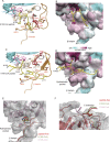
Figure 3. Substrate binding and conformational changes of the central β hairpin and C extension.
(A) Details of xEco2-K105-CoA interaction showing substrate hairpin (yellow) coordinated by the C extension (red) in form of a β sheet. (B) Conserved surface rendition of K105-CoA bound to xEco2. (C) Details of xEco2-K106-CoA interaction showing substrate peptide (yellow) coordinated by the central β hairpin (orange) in form of a β sheet. (D) Conserved surface rendition of K106-CoA bound to xEco2. (E) Superimposition of peptide-free (red), K105-CoA (green), and K106-CoA (salmon) bound xEco2 structures showing occupation of and displacement of W623 from the central hydrophobic pocket. (F) Superimposition of the C extension in peptide-free (red), K105-CoA (green), and K106-CoA (salmon) bound conformations. The C extension shows a 180° flip from coordinating W623 at the hydrophobic pocket to an open conformation upon K106-CoA binding.
Figure 4
Figure 4. Mechanism of Eco1-mediated Smc3 acetylation.
(A) Speculative model for Eco1-Smc3 interaction created by docking of xEco2-K105-CoA structure (salmon and blue) onto the S. cerevisiae Smc3-Scc1 (Smc3 is green; Scc1 is cyan) structure with reference to the relative positions of the tandem lysines (S. cerevisiae K112 and K113). (B) Speculative model for Eco1-Smc3 interaction created by docking of xEco2-K106-CoA structure (salmon and blue) onto the S. cerevisiae Smc3-Scc1 (Smc3 is green; Scc1 is cyan) structure with reference to the relative positions of the tandem lysines (S. cerevisiae K112 and K113). xEco2 rotates by 180° along the plane of the β sheet of Smc3 ATPase domain. (C) Schematics of Eco1 substrate specificity conferred by the concerted conformational changes of the central β hairpin (orange) and the C extension (red). The conserved hydrophobic pocket is occupied by W623 of xEco2 in the peptide-free and K105 targeting configurations, and by Y109 from Smc3 during K106 targeting respectively.

References

    1. Michaelis C., Ciosk R. & Nasmyth K. Cohesins: chromosomal proteins that prevent premature separation of sister chromatids. Cell 91, 35–45 (1997). - PubMed
    1. Guacci V., Koshland D. & Strunnikov A. A direct link between sister chromatid cohesion and chromosome condensation revealed through the analysis of MCD1 in S. cerevisiae. Cell 91, 47–57 (1997). - PMC - PubMed
    1. Losada A., Hirano M. & Hirano T. Identification of Xenopus SMC protein complexes required for sister chromatid cohesion. Genes Dev 12, 1986–1997 (1998). - PMC - PubMed
    1. Ciosk R. et al.. Cohesin’s binding to chromosomes depends on a separate complex consisting of Scc2 and Scc4 proteins. Mol Cell 5, 243–254 (2000). - PubMed
    1. Murayama Y. & Uhlmann F. Biochemical reconstitution of topological DNA binding by the cohesin ring. Nature 505, 367–371 (2014). - PMC - PubMed

Publication types

MeSH terms

LinkOut - more resources