

Value of second-opinion review of outside institution PET-CT examinations
- PMID: 28291158
- PMCID: PMC5354085
- DOI: 10.1097/MNM.0000000000000647
Value of second-opinion review of outside institution PET-CT examinations
Abstract
Objective: This study aimed to determine whether second-opinion reviews of PET/CT examinations by subspecialists alter reporting of malignant findings.
Materials and methods: This IRB-approved study compared 240 fluorine-18 fluorodeoxyglucose PET/CT consecutively dictated reports by two nuclear medicine subspecialists against the original outside institution reports. Subspecialist reviews documented whether malignant findings on the outside report were malignant and noted additional malignant findings not described on the outside report. The final diagnosis of malignancy or benignity was determined by pathology when available, otherwise by imaging follow-up.
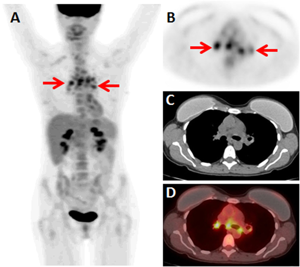
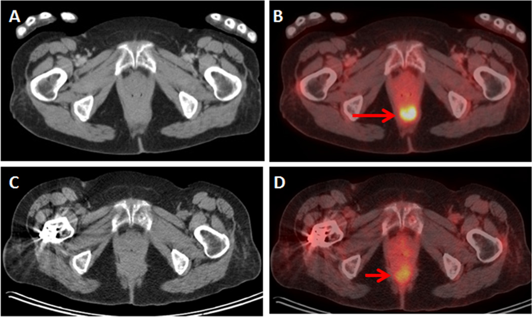
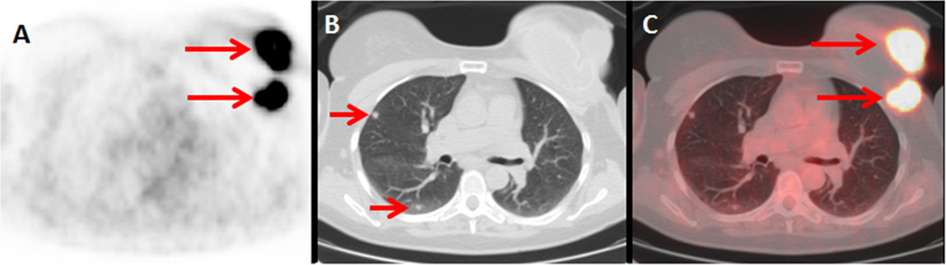
Results: A total of 22 findings (in 20 reports) called suspicious/malignant on the outside reports were deemed benign by subspecialist review. A final diagnosis was available for 20 of 22 findings by pathology (n=3) or follow-up imaging (n=17). The subspecialist review was accurate in 20 (100%) of 20 cases where a final diagnosis was available. The subspecialist review called 11 findings (in 11 reports) suspicious/malignant that were not described or deemed benign on the outside reports. Definitive diagnosis was available for 10 of 11 findings by pathology (n=7) or follow-up imaging (n=3). The second-opinion report was accurate in seven (70%) of 10 cases where a final diagnosis was available.
Conclusion: In 31 (13%) of 240 fluorine-18 fluorodeoxyglucose PET/CT examinations performed at an outside institution, subspecialist review resulted in at least one discordant opinion of malignancy. For 28 discrepant cases where a final diagnosis was available, the subspecialist review defined malignancy or benignity correctly in 25 (89%) of 28 cases. This provides evidence for the cost and effort invested in performing second-opinion reviews of PET/CT studies.
Figures
References
-
- Gollub MJ, Panicek DM, Bach AM, Penalver A, Castellino RA. Clinical importance of reinterpretation of body CT scans obtained elsewhere in patients referred for care at a tertiary cancer center. Radiology. 1999;210(1):109–112. - PubMed
-
- Loughrey GJ, Carrington BM, Anderson H, Dobson MJ, Lo Ying Ping F. The value of specialist oncological radiology review of cross-sectional imaging. Clin Radiol. 1999;54(3):149–154. discussion 154-145. - PubMed
-
- Leung JW, Margolin FR, Dee KE, Jacobs RP, Denny SR, Schrumpf JD. Performance parameters for screening and diagnostic mammography in a community practice: are there differences between specialists and general radiologists? AJR Am J Roentgenol. 2007;188(1):236–241. - PubMed
MeSH terms
Substances
Grants and funding
LinkOut - more resources
Full Text Sources
Other Literature Sources
