Review
. 2017 May;90(1073):20160920.
doi: 10.1259/bjr.20160920. Epub 2017 Apr 6.

Juvenile idiopathic arthritis: what is the utility of ultrasound?

Affiliations
Review

Juvenile idiopathic arthritis: what is the utility of ultrasound?

Hershernpal A S Basra et al. Br J Radiol. 2017 May.

Abstract

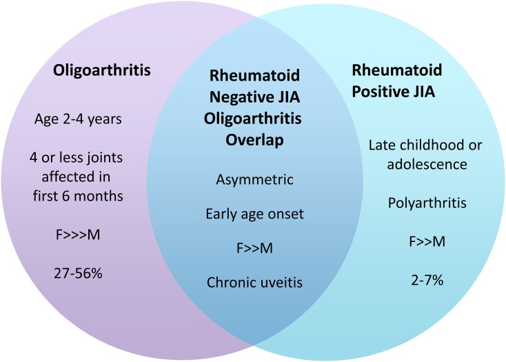
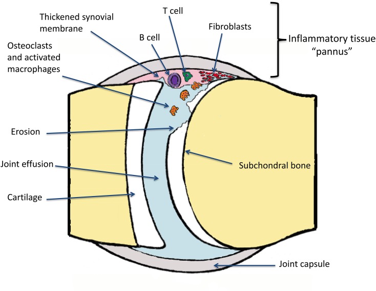
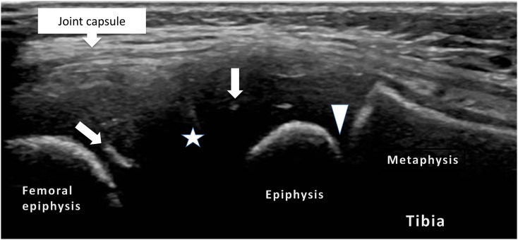
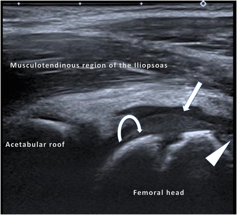
Juvenile idiopathic arthritis (JIA) is a heterogeneous condition and an important cause of acquired disability in children. Evidence supports early treatment to prevent future complications. This relies on prompt diagnosis, achieved by a high index of clinical suspicion and supportive evidence, including the detection of joint and or tendon inflammation. Ultrasound is a readily accessible, well-tolerated, safe and accurate modality for assessing joints and the surrounding soft tissues. It can also be used to guide therapy into those joints and tendon sheaths resistant to systemic treatments. Ultrasound imaging is highly operator dependent, and the developing skeleton poses unique challenges in interpretation with sonographic findings that can mimic pathology and vice versa. Ultrasound technology has been rapidly improving and is more accessible than ever before. In this article, we review the normal appearances, highlight potential pitfalls and present the key pathological findings commonly seen in JIA.


Figures

Figure 1.
Figure 1.
Heterogeneity within juvenile idiopathic arthritis (JIA) classifications: features of some of the disease subtypes can overlap with others, causing difficulties in accurate classification. This has implications on diagnosis, treatment and follow-up. F, female; M, male.
Figure 2.
Figure 2.
Pathogenesis of joint damage: an unknown inciting trigger activates T and B cells of the adaptive immune system. These immune orchestrators promote joint infiltration by activating macrophages and promoting osteoclastic activity. Over time, fibroblast and chondrocyte functions are altered, impairing healing. The synovium becomes thickened and the cartilage is degraded, exposing the subchondral bone to further erosive damage. Fluid can collect in the joint forming an effusion.
Figure 3.
Figure 3.
Lateral, longitudinal views of a normal knee joint: the image shows the potential joint space (star) lined by the hyaline cartilage and linear and radial echoes (arrows) representing vascular channels within the epiphyseal cartilage and the physeal cartilage (arrowhead).
Figure 4.
Figure 4.
An anterior longitudinal image of the suprapatella recess of the knee: active nodular synovitis (double-headed arrow) in the suprapatella recess and synovial thickening (arrowhead) in the visualized joint is shown. An anechoic joint effusion (x) is seen above the prefemoral fat pad (star), which was also hyperaemic (not shown). The unfused physis (arrow) is also demonstrated in this child with juvenile idiopathic arthritis.
Figure 5.
Figure 5.
An anterior longitudinal image of the hip joint: a mildly echogenic normal epiphyseal cartilage (curved arrow) is shown. When this is compared with the adjacent anechoic joint effusion (arrow), synovial proliferation is just visible at the femoral head–neck junction (arrowhead), which is similar in echogenicity to the unossified cartilage. Power Doppler interrogation of the synovium (not included) did not show any active synovitis.
Figure 6.
Figure 6.
Axial views of the medial ankle tendons in a patient with juvenile idiopathic arthritis: the tibialis posterior tendon (arrowhead) surrounded by hypoechoic fluid (arrow) and mildly echogenic tenosynovitis (curved arrows) (a) and the power Doppler signal in keeping with active tenosynovitis (b).
Figure 7.
Figure 7.
Longitudinal (a) and axial (b) images of a damaged third and normal fourth metacarpophalangeal joint in a patient with juvenile idiopathic arthritis: the preserved chondroepiphysis and hyaline cartilage (star) as compared with the deformed third metacarpal head, which contains erosions (arrows), synovial thickening/pannus (curved arrows) and oedema in the soft tissues (thick double-headed arrow), blurring boundaries between the normal tissue layers (thin double-sided arrow), can be noted.

Similar articles

Cited by

References

    1. Thierry S, Fautrel B, Lemelle I, Guillemin F. Prevalence and incidence of juvenile idiopathic arthritis: a systematic review. Joint Bone Spine 2014; 81: 112–17. doi: https://doi.org/10.1016/j.jbspin.2013.09.003 - DOI - PubMed
    1. Petty RE, Southwood TR, Manners P, Baum J, Glass DN, He X, et al. . International league of associations for rheumatology classification of juvenile idiopathic arthritis: second revision, Edmonton, 2001; 31. - PubMed
    1. Rigante D, Bosco A, Esposito S. The etiology of juvenile idiopathic arthritis. Clin Rev Allergy Immunol 2015; 49: 253–61. doi: https://doi.org/10.1007/s12016-014-8460-9 - DOI - PubMed
    1. Burgos-Vargas R. Juvenile onset spondyloarthropathies: therapeutic aspects. Ann Rheum Dis 2002; 61(Suppl. 3): 33–9. - PMC - PubMed
    1. Martini A. Are the number of joints involved or the presence of psoriasis still useful tools to identify homogeneous disease entities in juvenile idiopathic arthritis? J Rheumatol 2003; 30: 1900–3. - PubMed