. 2017 Mar 1;73(Pt 3):138-145.
doi: 10.1107/S2053230X17002102. Epub 2017 Feb 21.

Proteolysis of truncated hemolysin A yields a stable dimerization interface

Affiliations

Proteolysis of truncated hemolysin A yields a stable dimerization interface

Walter R P Novak et al. Acta Crystallogr F Struct Biol Commun. .

Abstract

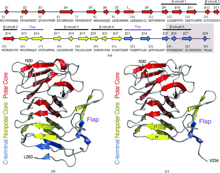
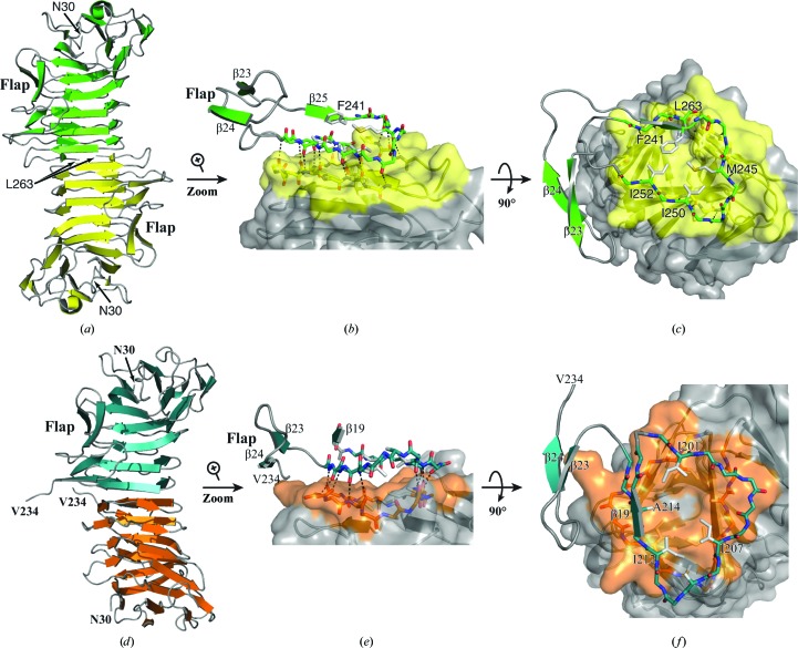
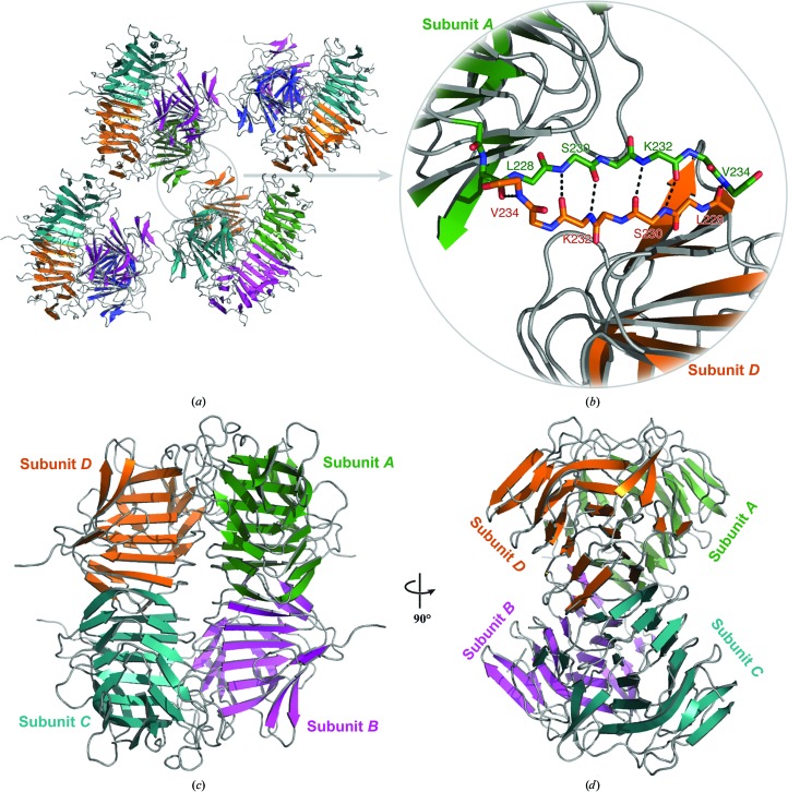
Wild-type and variant forms of HpmA265 (truncated hemolysin A) from Proteus mirabilis reveal a right-handed, parallel β-helix capped and flanked by segments of antiparallel β-strands. The low-salt crystal structures form a dimeric structure via the implementation of on-edge main-chain hydrogen bonds donated by residues 243-263 of adjacent monomers. Surprisingly, in the high-salt structures of two variants, Y134A and Q125A-Y134A, a new dimeric interface is formed via main-chain hydrogen bonds donated by residues 203-215 of adjacent monomers, and a previously unobserved tetramer is formed. In addition, an eight-stranded antiparallel β-sheet is formed from the flap regions of crystallographically related monomers in the high-salt structures. This new interface is possible owing to additional proteolysis of these variants after Tyr240. The interface formed in the high-salt crystal forms of hemolysin A variants may mimic the on-edge β-strand positioning used in template-assisted hemolytic activity.

Keywords: Proteus mirabilis; alternate crystal forms; hemolysin A; proteolysis; two-partner secretion; β-helix.


Figures

Figure 1
Figure 1
(a) Secondary-structure diagram of HpmA265. The gray box indicates the region removed by proteolysis in the high-salt structure. The cartoon representations denote the subdomain architecture embedded within (b) the low-salt HpmA265 structure and (c) the high-salt HpmA265 structure. Despite proteolysis in the high-salt structure, both the low-salt and high-salt structures harbor polar core, nonpolar core and carboxy-terminal subdomains. The β-helix core of the HpmA265 structure comprises both the polar and nonpolar core subdomains. Additionally, a four-stranded antiparallel β-sheet (flap) frames one side of the β-helix core. The low-salt crystal form (b) includes residues Asn30–Leu263 and extends through β28 at the C-terminus. The high-salt form (c) includes residues Asn30–Val234 and terminates at β24, which is the final antiparallel β-strand within the flap region.
Figure 2
Figure 2
2F oF c electron-density maps contoured at 1σ near the mutation site. (a) shows the low-salt HpmA265 structure in green. (b) shows the low-salt Y134A structure in magenta and clear density for two ordered water molecules in place of the tyrosine side chain. (c) shows the low-salt AA double-mutant structure in cyan. Despite the loss of these two side chains the overall structure remains unaffected, and no electron density is observed that may account for ordered water molecules. Fig. 2 was created with the UCSF Chimera molecular visualization program (Pettersen et al., 2004 ▸).
Figure 3
Figure 3
Dimerization interfaces in the low-salt and high-salt HpmA265 structures. (a) The HpmA265 low-salt crystal structure uses β26–β28 (residues Ser244–Leu263) at the dimerization interface. (b) Magnification of the side view showing hydrogen bonds donated by exposed on-edge β-strands (β26, β27 and β28 residues) from each monomer in the low-salt structure. (c) Top view emphasizing van der Waals interactions during the formation of the low-salt dimer interface. (d) In the AA high-salt crystal structure, proteolysis allows β20–β22 (residues Arg200–Phe215) to establish the dimer interface. (e) Side view emphasizing the hydrogen bonds donated by exposed on-edge β-strands (β20–β22, residues Arg200–Phe215) from each monomer in the high-salt AA structure. (f) A top view showing the van der Waals interactions of Ile201, Ile207, Ile212 and Ala214 during the formation of the high-salt dimer interface.
Figure 4
Figure 4
Tetramer formation in the high-salt structures. (a) Depiction of the eight-stranded antiparallel β-sheet formed from the flap regions of crystallographically related tetramers in the high-salt condition. The removal of β25–β28 owing to the proteolysis event has allowed β24 to expose on-edge main-chain hydrogen-bonding partners. (b) Magnified view of the main-chain atoms, including those donated from β24, used to assemble the inter-subunit antiparallel β-sheet. The newly discovered dimerization facilitates the formation of a stable tetramer (c, d). Interactions between adjacent N-­terminal polar core subdomains form the majority of this interface.

Similar articles

Cited by

References

    1. Adams, P. D. et al. (2010). Acta Cryst. D66, 213–221. - PubMed
    1. Baelen, S., Dewitte, F., Clantin, B. & Villeret, V. (2013). Acta Cryst. F69, 1322–1327. - PMC - PubMed
    1. Chen, V. B., Arendall, W. B., Headd, J. J., Keedy, D. A., Immormino, R. M., Kapral, G. J., Murray, L. W., Richardson, J. S. & Richardson, D. C. (2010). Acta Cryst. D66, 12–21. - PMC - PubMed
    1. Choi, J. H., May, B. C. H., Govaerts, C. & Cohen, F. E. (2009). Structure, 17, 1014–1023. - PubMed
    1. Clantin, B., Hodak, H., Willery, E., Locht, C., Jacob-Dubuisson, F. & Villeret, V. (2004). Proc. Natl Acad. Sci. USA, 101, 6194–6199. - PMC - PubMed

MeSH terms