. 2017 Mar 1;73(Pt 3):167-173.
doi: 10.1107/S2053230X17001376. Epub 2017 Feb 28.

Crystal structure of the type IV secretion system component CagX from Helicobacter pylori

Affiliations

Crystal structure of the type IV secretion system component CagX from Helicobacter pylori

Jin Zhang et al. Acta Crystallogr F Struct Biol Commun. .

Abstract

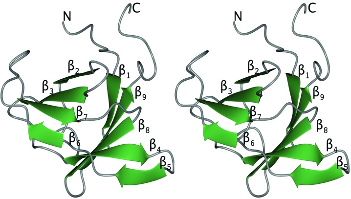
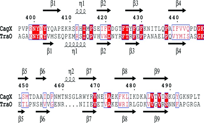
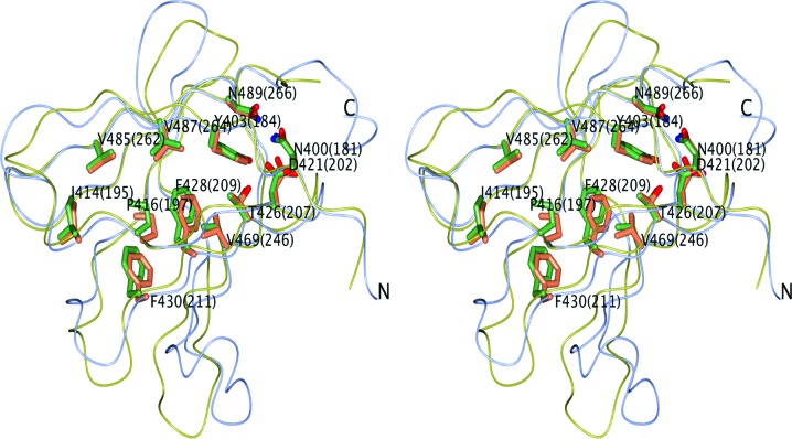
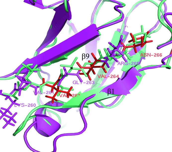
Helicobacter pylori, a Gram-negative bacterial pathogen prevalent in the human population, is the causative agent of severe gastric diseases. An H. pylori type IV secretion (T4S) system encoded by the cytotoxin-associated gene pathogenicity island (cagPAI) is responsible for communication with host cells. As a component of the cagPAI T4S system core complex, CagX plays an important role in virulence-protein translocation into the host cells. In this work, the crystal structure of the C-terminal domain of CagX (CagXct), which is a homologue of the VirB9 protein from the VirB/D4 T4S system, is presented. CagXct is only the second three-dimensional structure to be elucidated of a VirB9-like protein. Another homologue, TraO, which is encoded on the Escherichia coli conjugative plasmid pKM101, shares only 19% sequence identity with CagXct; however, there is a remarkable similarity in tertiary structure between these two β-sandwich protein domains. Most of the residues that are conserved between CagXct and TraO are located within the protein core and appear to be responsible for the preservation of this domain fold. The studies presented here will contribute to our understanding of different bacterial T4S systems.

Keywords: CagX; Helicobacter pylori; X-ray diffraction; cagPAI; crystal structure; cytotoxin-associated gene pathogenicity island; type IV secretion system.


Figures

Figure 1
Figure 1
15% SDS–PAGE analysis of purified CagXct stained with Coomassie Brilliant Blue. Lane A, non-induced expression strains. Lane a, induced expression strains. Lane B, pellet fractions. Lane b, supernatant fractions. Lane 1, eluted with 500 mM imidazole; the band containing CagXct with a Trx tag is around 35 kDa. Lane 2, after cleavage by TEV; the band containing CagXct is under 14.4 kDa. Lane 3, flowthrough of the second nickel column. Lane 4, the sample before size-exclusion chromatography, showing high purity. Lane M contains molecular-mass marker (labelled in kDa).
Figure 2
Figure 2
(a) Size-exclusion chromatography trace (HiLoad 16/600 Superdex 200 75 pg; GE Healthcare). CagXct elutes at 80–110 ml, which is in agreement with its monomer molecular mass of 11.4 kDa. The peak intensity is 192 mAU. Samples between 80 and 110 ml labelled 3–17 in red were picked for 15% SDS–PAGE analysis. (b) 15% SDS–PAGE analysis of the purified CagXct samples labelled 3, 5, 9, 11, 13, 15 and 17 in (a). Lane M contains molecular-mass marker (labelled in kDa).
Figure 3
Figure 3
Electron-density maps around β-sheet 2 of CagXct monomer A. The 2F o − F c map (blue) is contoured at 1.4σ and the F oF c map is contoured at 3.0σ (green) and −3.0σ (red). The difference density ripples (red and green) are likely to be owing to crystal twinning. This figure was prepared using PyMOL (Schrödinger).
Figure 4
Figure 4
A stereo diagram showing a cartoon representation of CagXct, with the β-strands coloured green and loops coloured grey. The secondary-structure elements of this β-sandwich domain are numbered. Figs. 4 and 6 were prepared using CCP4mg (McNicholas et al., 2011 ▸).
Figure 5
Figure 5
Anmino-acid sequence alignment of CagXct and TraOct. The secondary-structure elements are indicated above and below the alignment, respectively, as β-strands and η-helices (310-helices). Conserved residues are shown in red boxes; matching amino acids with similar properties are shown in blue boxes. The secondary-structure assignments were carried out and the figure was produced using ESPript3 (Robert & Gouet, 2014 ▸).
Figure 6
Figure 6
A stereo diagram showing the structural superposition of CagX (ice-blue worm model) and TraO (yellow; PDB entry 3jqo). Side chains of residues forming the conserved protein core are shown with C atoms in coral (CagX) or green (TraO). Residue numbers of TraO are given in parentheses after these of the CagX residues.
Figure 7
Figure 7
Comparison of the protein-binding region of the β9 strand (shown as sticks; β1 strands are shown as cartoons) in CagXct (cyan) and in TraO (magenta, only the residues of TraO are labelled). Apart from the binding residues, there is a high sequence-similarity pattern: ‘xVxVxN’ (these residues are shown in red; x represents the binding amino-acid residues).

Similar articles

Cited by

References

    1. Akopyants, N. S., Clifton, S., Kersulyte, D., Crabtree, J. E., Youree, B. E., Reece, C. A., Bukanov, N. O., Drazek, E. S., Roe, B. A. & Berg, D. E. (1998). Mol. Microbiol. 28, 37–53. - PubMed
    1. Backert, S., Churin, Y. & Meyer, T. F. (2002). Keio J. Med. 51, Suppl. 2, 6–14. - PubMed
    1. Backert, S. & Selbach, M. (2008). Cell. Microbiol. 10, 1573–1581. - PubMed
    1. Bayliss, R., Harris, R., Coutte, L., Monier, A., Fronzes, R., Christie, P. J., Driscoll, P. C. & Waksman, G. (2007). Proc. Natl Acad. Sci. USA, 104, 1673–1678. - PMC - PubMed
    1. Blaser, M. J. (1997). J. Clin. Invest. 100, 759–762. - PMC - PubMed

MeSH terms