. 2017 Mar 1;73(Pt 3):223-233.
doi: 10.1107/S2059798317001061. Epub 2017 Feb 22.

CheckMyMetal: a macromolecular metal-binding validation tool

Affiliations

CheckMyMetal: a macromolecular metal-binding validation tool

Heping Zheng et al. Acta Crystallogr D Struct Biol. .

Abstract

Metals are essential in many biological processes, and metal ions are modeled in roughly 40% of the macromolecular structures in the Protein Data Bank (PDB). However, a significant fraction of these structures contain poorly modeled metal-binding sites. CheckMyMetal (CMM) is an easy-to-use metal-binding site validation server for macromolecules that is freely available at http://csgid.org/csgid/metal_sites. The CMM server can detect incorrect metal assignments as well as geometrical and other irregularities in the metal-binding sites. Guidelines for metal-site modeling and validation in macromolecules are illustrated by several practical examples grouped by the type of metal. These examples show CMM users (and crystallographers in general) problems they may encounter during the modeling of a specific metal ion.

Keywords: CheckMyMetal; coordination geometry; metal-binding environment; validation.


Figures

Figure 1
Figure 1
Zinc-binding site in serum albumin, determined using X-rays above and below the zinc absorption edge. Gray, 2F oF c; orange, F oF c.
Figure 2
Figure 2
Mg998 in the CWP6 protein from the CWB2 cell-wall-anchoring module of the C. difficile cell-wall proteins CWP8 and CWP6
Figure 3
Figure 3
A water-binding site in the structure of proteinase K (PDB entry 3i34) which may be better interpreted as potassium.
Figure 4
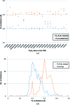
Figure 4
(a) Distribution of distances from iron to sulfur in an Fe2S2 cluster. For each Fe2S2 cluster (identified by CSD Refcode) the distances to all four sulfurs within the Fe2S2 cluster are shown in blue, while the distances to all four external sulfurs are shown in orange. (b) Distribution of Fe—S distances within Fe2S2 modeled in the PDB high-resolution data (<1.5 Å). Typical Fe—S distances are ∼2.2 Å between Fe and sulfurs within the Fe2S2 cluster, while typical Fe—S distances are ∼2.3 Å between Fe and external (cysteine) sulfurs.

Similar articles

Cited by

References

    1. Arcovito, A. & della Longa, S. (2012). J. Inorg. Biochem. 112, 93–99. - PubMed
    1. Ascone, I. & Strange, R. (2009). J. Synchrotron Rad. 16, 413–421. - PubMed
    1. Avvaru, B. S., Kim, C. U., Sippel, K. H., Gruner, S. M., Agbandje-McKenna, M., Silverman, D. N. & McKenna, R. (2010). Biochemistry, 49, 249–251. - PMC - PubMed
    1. Barber, J., Morris, E. & Büchel, C. (2000). Biochim. Biophys. Acta, 1459, 239–247. - PubMed
    1. Boal, A. K. & Rosenzweig, A. C. (2009). J. Am. Chem. Soc. 131, 14196–14197. - PMC - PubMed

Publication types