. 2017 Mar 14;15(3):e2000735.
doi: 10.1371/journal.pbio.2000735. eCollection 2017 Mar.

Three-dimensional preservation of cellular and subcellular structures suggests 1.6 billion-year-old crown-group red algae

Affiliations

Three-dimensional preservation of cellular and subcellular structures suggests 1.6 billion-year-old crown-group red algae

Stefan Bengtson et al. PLoS Biol. .

Abstract

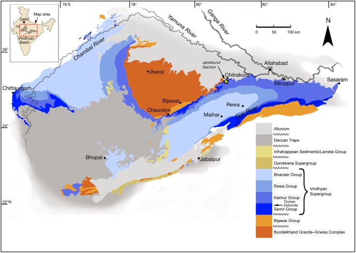
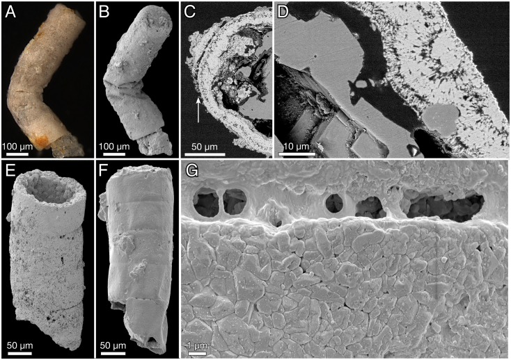
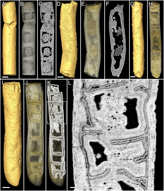
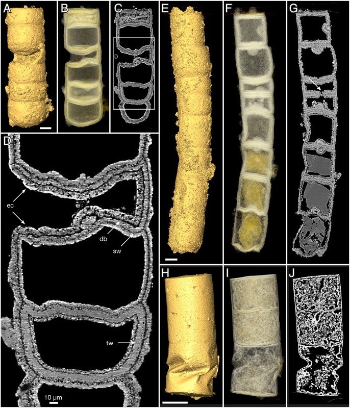
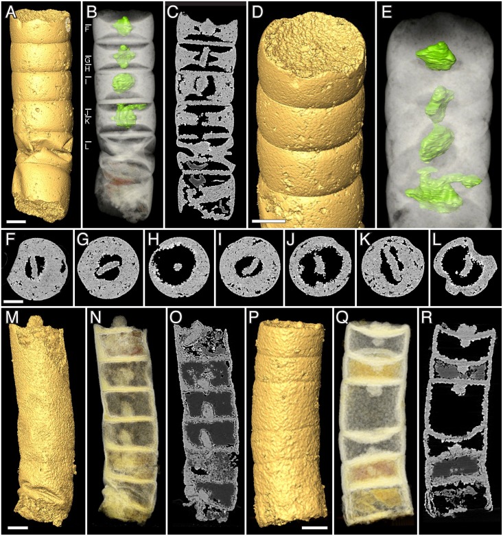
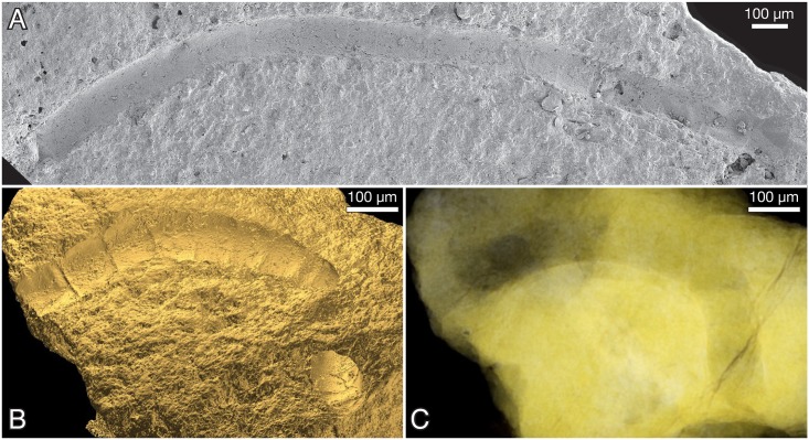
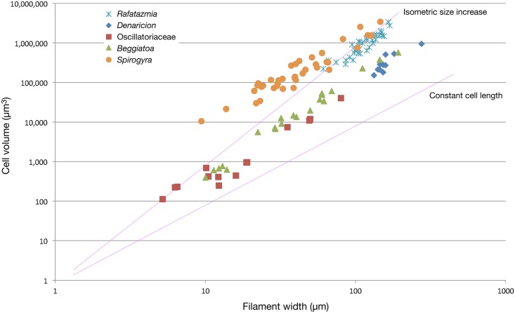
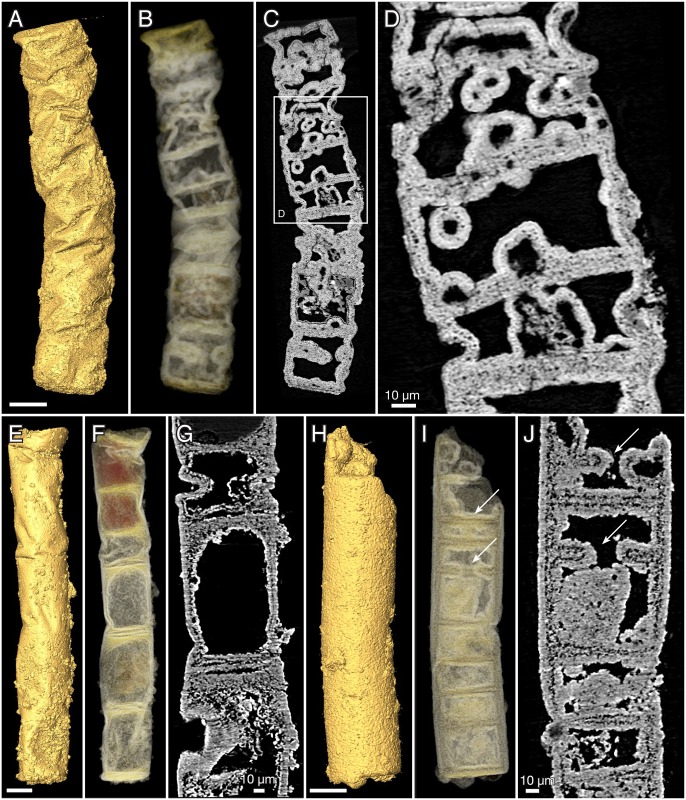
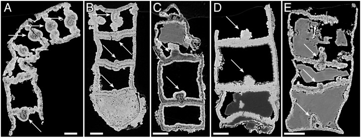
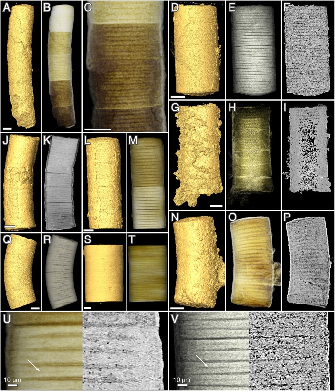
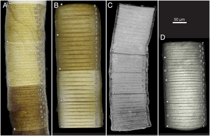
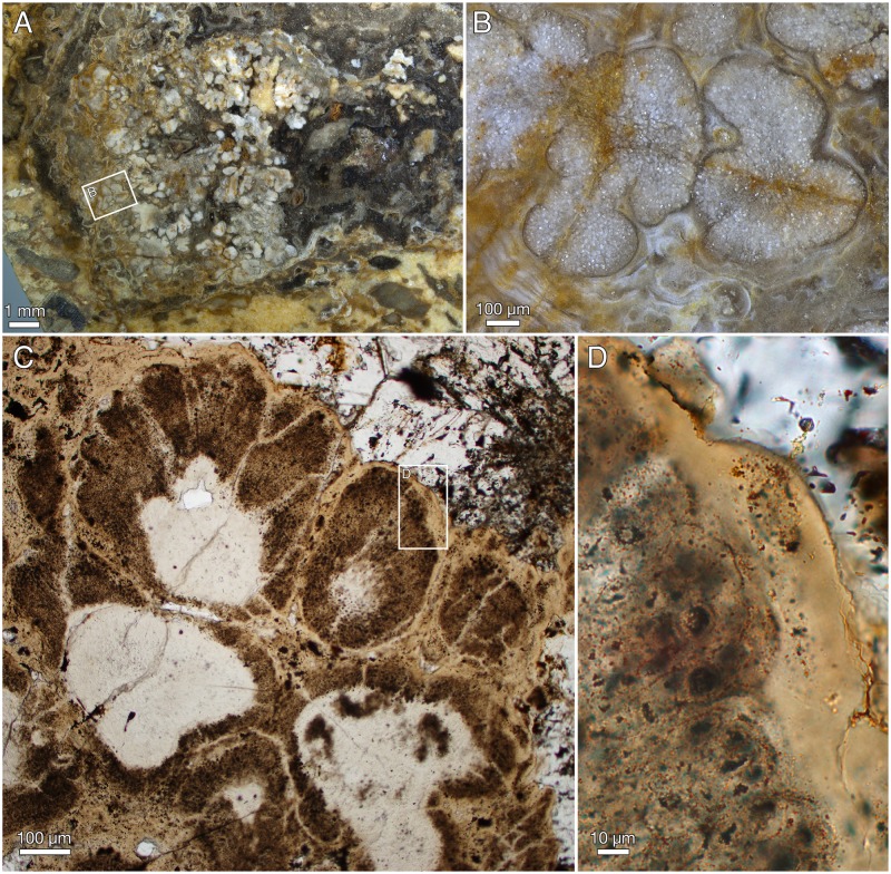
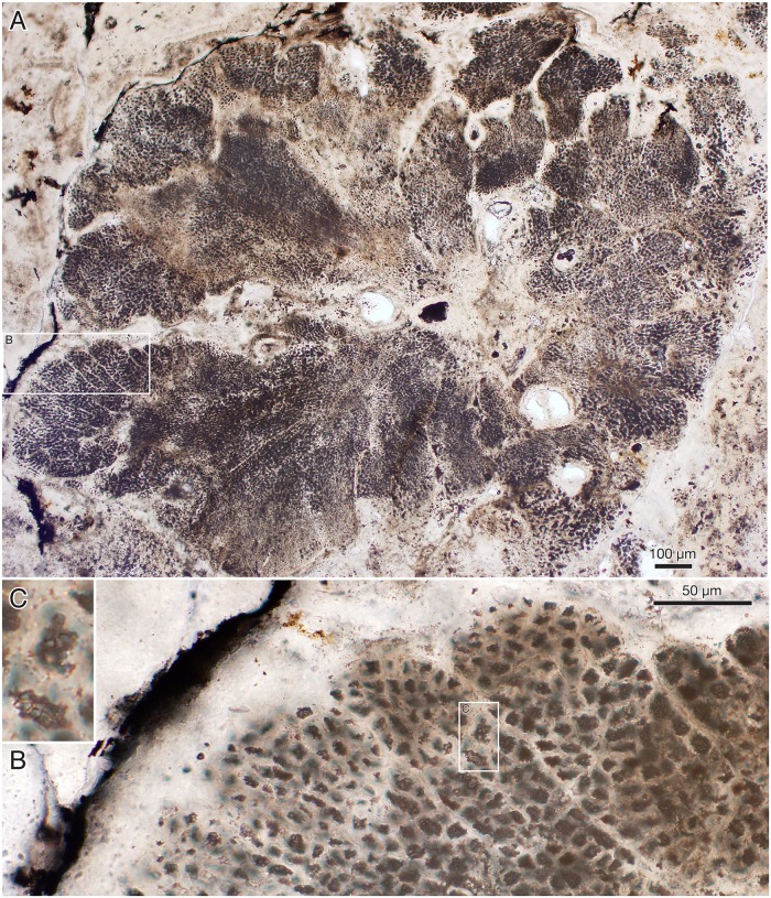
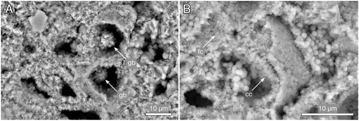
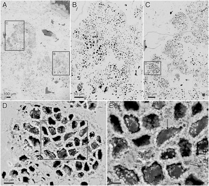
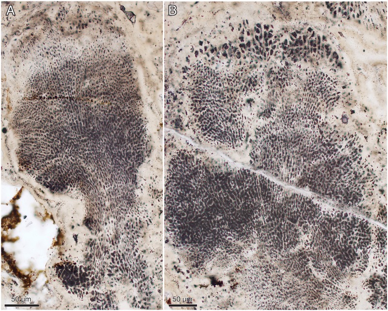
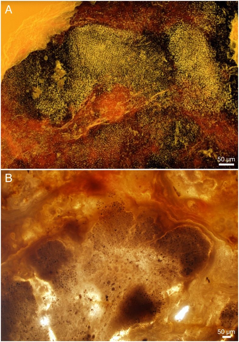
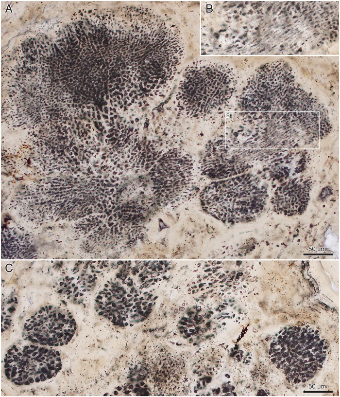
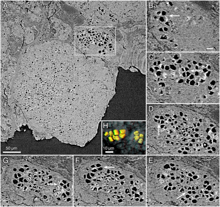
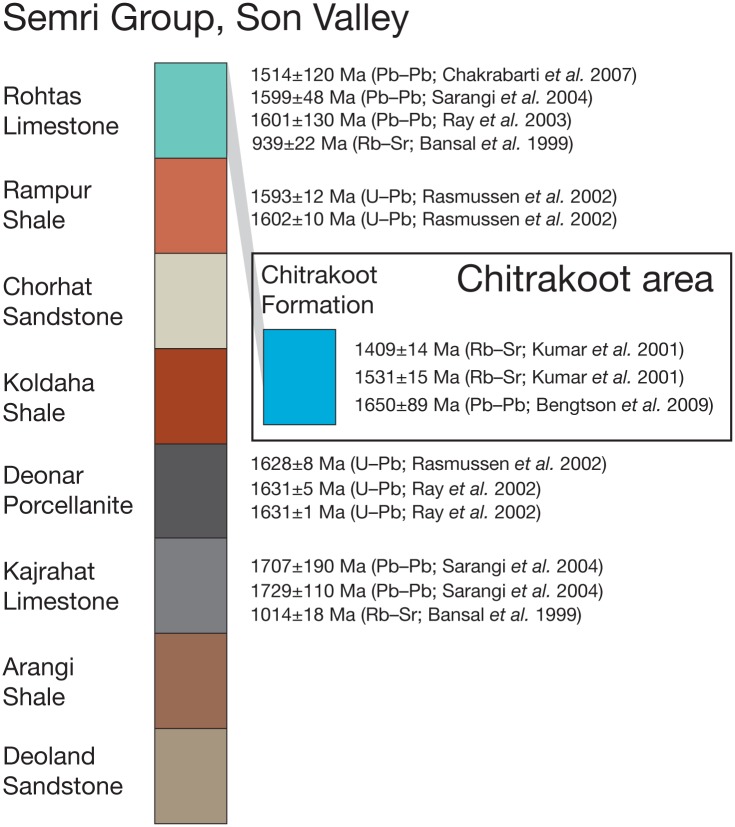
The ~1.6 Ga Tirohan Dolomite of the Lower Vindhyan in central India contains phosphatized stromatolitic microbialites. We report from there uniquely well-preserved fossils interpreted as probable crown-group rhodophytes (red algae). The filamentous form Rafatazmia chitrakootensis n. gen, n. sp. has uniserial rows of large cells and grows through diffusely distributed septation. Each cell has a centrally suspended, conspicuous rhomboidal disk interpreted as a pyrenoid. The septa between the cells have central structures that may represent pit connections and pit plugs. Another filamentous form, Denaricion mendax n. gen., n. sp., has coin-like cells reminiscent of those in large sulfur-oxidizing bacteria but much more recalcitrant than the liquid-vacuole-filled cells of the latter. There are also resemblances with oscillatoriacean cyanobacteria, although cell volumes in the latter are much smaller. The wider affinities of Denaricion are uncertain. Ramathallus lobatus n. gen., n. sp. is a lobate sessile alga with pseudoparenchymatous thallus, "cell fountains," and apical growth, suggesting florideophycean affinity. If these inferences are correct, Rafatazmia and Ramathallus represent crown-group multicellular rhodophytes, antedating the oldest previously accepted red alga in the fossil record by about 400 million years.


Conflict of interest statement

The authors have declared that no competing interests exist.

Figures

Fig 1
Fig 1. Geological map of the Vindhyan basin, central India.
After Azmi et al. [55], based on several sources.
Fig 2
Fig 2. Rafatazmia chitrakootensis n. gen., n. sp., Scanning Electron Microscopy (SEM) images.
(A–D) NRM S156424 (also figured in [52]); C and D are backscatter images of a polished transverse section. (E) NRM X5647. (F, G) NRM X5648.
Fig 3
Fig 3. Rafatazmia chitrakootensis n. gen., n. sp., Synchrotron-Radiation X-ray Tomographic Microscopy (SRXTM) renderings.
(A–C) NRM X5646, surface, volume, slice. (D–F) NRM X5592, surface, volume, slice. (G, H) NRM X4230, surface, volume. (I–L) NRM X4229, surface, volume, slice. Legend: sw, septal wall; is, incomplete septum. Scale bars 50 μm.
Fig 4
Fig 4. Rafatazmia chitrakootensis n. gen., n. sp., SRXTM renderings.
(A–D) NRM X4251, surface, volume, slice. (E–G) NRM X5572, surface, volume, slice. (H–J) NRM X5562, surface, volume, slice. Legend: ec, external coating; db, diagenetic boundary; sw, septal wall; tw, thick wall. Scale bars 50 μm except where otherwise noted.
Fig 5
Fig 5. Rafatazmia chitrakootensis n. gen., n. sp., SRXTM renderings.
(A–L) Holotype, NRM X4258. (A) Surface rendering. (B) Volume rendering with rhomboidal disks coloured for visibility. (C) Virtual slice. (D) Surface. (E) Volume. (F–L) Transverse slices (positions indicated in B). (M–O) NRM X5620, surface, volume, slice. (P–R) NRM X5574, surface, volume, slice. Scale bars 50 μm.
Fig 6
Fig 6. Rafatazmia chitrakootensis n. gen., n. sp., SEM image (A) and SRXTM renderings (B, C).
A: NRM S156422 (also figured in [52]). (B, C) NRM X5544, surface, volume.
Fig 7
Fig 7. Relationships between filament width and cell volume in Rafatazmia, Denaricion, and some modern filamentous organisms.
Data sources for the modern taxa: Oscillatoriacea [–64] and Soft-Bodied Stream Algae of California (http://dbmuseblade.colorado.edu/DiatomTwo/sbsac_site/). Beggiatoa [–70]. Spirogyra (Soft-Bodied Stream Algae of California). Individual measurements are in Supporting Information in the file S1 Data.
Fig 8
Fig 8. Rafatazmia chitrakootensis n. gen., n. sp., SRXTM renderings.
(A–D) NRM X4246, surface, volume, slice. (E–G) NRM X4240, surface, volume, slice. (H–J) NRM X4242, surface, volume, slice; arrows point to new septa with central pore. Scale bars 50 μm except where otherwise noted.
Fig 9
Fig 9. Rafatazmia chitrakootensis n. gen., n. sp., SRXTM virtual slices.
Arrows point to diagenetic overgrowths indicating the presence of irregularities consistently placed at the centre of septa and interpreted as possible pit plugs. (A) NRM X5573. (B) NRM X5645. (C) NRM X5626. (D) NRM X5610. (E) NRM X5613. Scale bars 50 μm.
Fig 10
Fig 10. Denaricion mendax n. gen., n. sp., SRXTM renderings.
(A–C) Holotype, NRM X5644, surface, volume, slice. (D–F, V) NRM X5604, surface, volume, slice, volume/slice. (G–I) NRM X5634, surface, volume, slice. (J, K) NRM X4233, surface, volume. (L, M, U) NRM X4234, surface, volume, volume/slice. (N–P) NRM X4244, surface, volume, slice. (Q, R) NRM X4256, surface, volume. (S–T) NRM X4235, surface, volume. Arrows in U and V point to dark lines suggesting incipient septation. Scale bars 50 μm except where otherwise noted.
Fig 11
Fig 11. Denaricion mendax n. gen., n. sp., blowups of SRXTM volume renderings from Fig 10 to show 4/8/16 pattern of dividing cells within compartments.
Asterisks show divisions between 8-cell compartments; rings mark weaker divisions between the fourth and the fifth cell within some compartments. (A) Same as in Fig 10B. (B) Same as in Fig 10M. (C) Same as in Fig 10K. (D) Same as in Fig 10E.
Fig 12
Fig 12. Ramathallus lobatus n. gen., n. sp.
(A, B) NRM X5639, reflected light. (C, D) NRM X5643, thin section.
Fig 13
Fig 13. Ramathallus lobatus n. gen., n. sp., holotype, NRM X5638.
Thin section. (A) Overview of specimen. (B) Detail of A to show cell structure in finger-like protrusions and noncellular apatitic coating. (C) Detail of B to show dark granular material within cells.
Fig 14
Fig 14. Ramathallus lobatus n. gen., n. sp., NRM X5640.
Environmental scanning electron microscopy (ESEM) images, backscatter mode. (A) Globular bodies (gb) within cells. (B) Coarse crystals (cc) and fine crystals (fc) in diagenetically phosphatized cell walls.
Fig 15
Fig 15. Ramathallus lobatus n. gen., n. sp., NRM X5638.
ESEM images, backscatter mode. (A) Overview. (B and C) Details of A to show pseudoparenchymatous structure with “cell fountains” and varying cell sizes. (D and E) Details of C to show preservation of cells. Legend: cm, carbonaceous matter; gm, granular matter.
Fig 16
Fig 16. Ramathallus lobatus n. gen., n. sp., NRM X5638.
Thin section. (A) Thallus with well-developed “cell fountains” in club-like distal part. (B) Thallus with “cell fountains” and cortex-like structure of enlarged cells.
Fig 17
Fig 17. Ramathallus lobatus n. gen., n. sp.
(A) NRM X5641, SRXTM volume rendering. (B) NRM X5642, thick section.
Fig 18
Fig 18. Ramathallus lobatus n. gen., n. sp., NRM X5638.
Thin section. (A) Overview of complex thallus with cell fountains. (B) Detail of A to show elongated cells. (C) Different area to A of the same thin section, showing globular or finger-shaped protrusions.
Fig 19
Fig 19. Ramathallus lobatus n. gen., n. sp., NRM X5641.
SRXTM slices. (A) Larger field of view to show the location of the imaged volume. (B–G) Consecutive sections through volume of larger cells, where putative tetraspores are marked with arrows. (H) Stereo anaglyph showing surface-rendered cell volumes of three putative tetraspores (yellow) surrounded by nontetradially arranged cells (grey, transparent).
Fig 20
Fig 20. Summary of recent published results from radiometric age determinations of Lower Vindhyan rocks in the Son Valley and the Chitrakoot area.
The shaded band indicates correlation of the Tirohan Dolomite (Chitrakoot Formation) with the Rohtas Limestone. Uncertainties are 2σ.

References

    1. Butterfield NJ. Animals and the invention of the Phanerozoic Earth system. Trends in Ecology and Evolution. 2011;26(2):81–7. 10.1016/j.tree.2010.11.012 - DOI - PubMed
    1. James TY, Kauff F, Schoch CL, Matheny PB, Hofstetter V, Cox CJ, et al. Reconstructing the early evolution of Fungi using a six-gene phylogeny. Nature. 2006;443(7113):818–22. 10.1038/nature05110 - DOI - PubMed
    1. Lücking R, Huhndorf S, Pfister DH, Plata ER, Lumbsch HT. Fungi evolved right on track. Mycologia. 2009;101(6):810–22. - PubMed
    1. Taylor TN, Krings M, Taylor EL. Fossil Fungi. Amsterdam: Elsevier; 2015.
    1. Xiao S. Written in stone: the fossil record of early eukaryotes In: Trueba G, Montúfar C, editors. Evolution from the Galapagos, Social and Ecological Interactions in the Galapagos Islands 2 New York: Springer; 2013. p. 107–24.

Publication types