Identification of cerebral perfusion using arterial spin labeling in patients with seizures in acute settings
- PMID: 28291816
- PMCID: PMC5349669
- DOI: 10.1371/journal.pone.0173538
Identification of cerebral perfusion using arterial spin labeling in patients with seizures in acute settings
Abstract
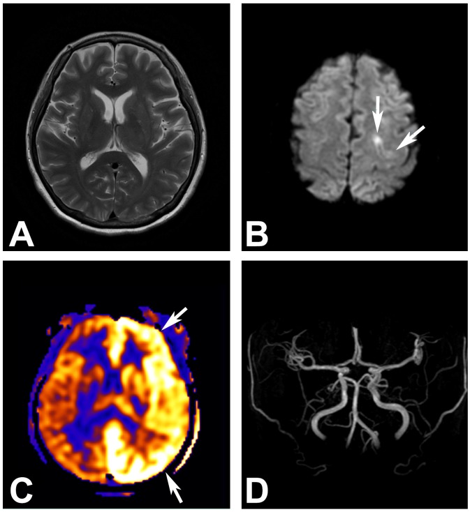
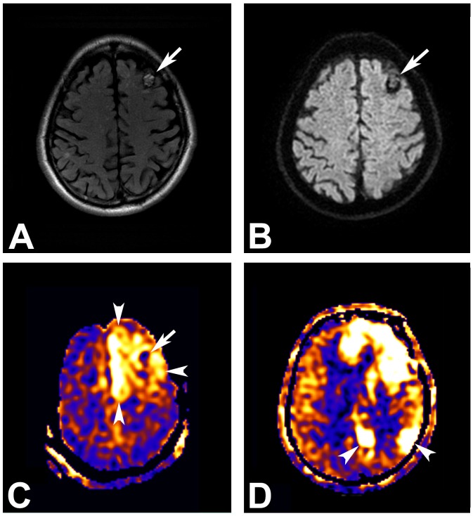
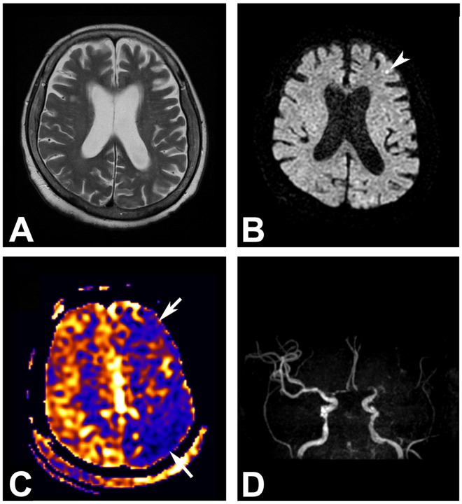
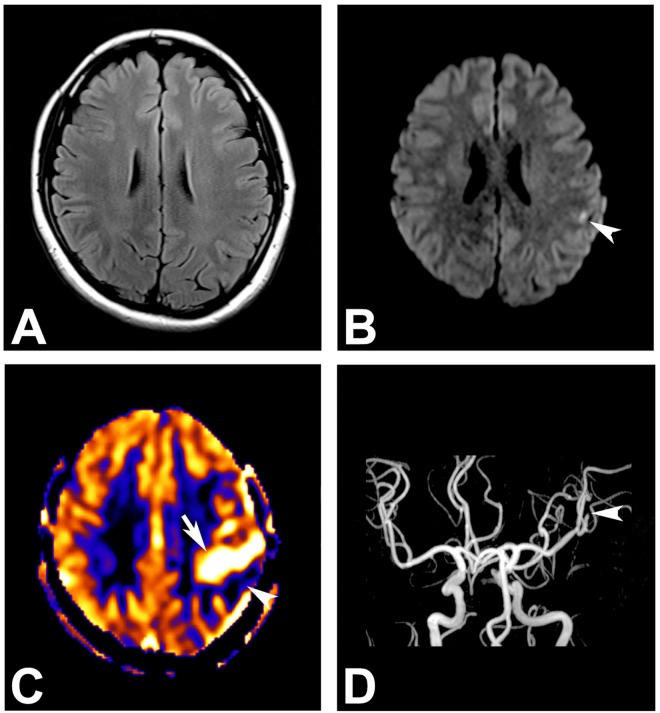
This study aimed to explore the utility of arterial spin labeling perfusion-weighted imaging (ASL-PWI) in patients with suspected seizures in acute settings. A total of 164 patients who underwent ASL-PWI for suspected seizures in acute settings (with final diagnoses of seizure [n = 129], poststroke seizure [n = 18], and seizure mimickers [n = 17]), were included in this retrospective study. Perfusion abnormality was analyzed for: (1) pattern, (2) multifocality, and (3) atypical distribution against vascular territories. Perfusion abnormality was detected in 39% (50/129) of the seizure patients, most (94%, 47/50) being the hyperperfusion pattern. Of the patients with perfusion abnormality, multifocality or hemispheric involvement and atypical distribution against vascular territory were revealed in 46% (23/50) and 98% (49/50), respectively. In addition, seizures showed characteristic features including hyperperfusion (with or without non-territorial distribution) on ASL-PWI, thus differentiating them from poststroke seizures or seizure mimickers. In patients in whom seizure focus could be localized on both EEG and ASL-PWI, the concordance rate was 77%. The present study demonstrates that ASL-PWI can provide information regarding cerebral perfusion status in patients with seizures in acute settings and has the potential to be used as a non-invasive imaging tool to identify the cerebral perfusion in patients with seizures.
Conflict of interest statement
Figures


Similar articles
-
Capability of arterial spin labeling MR imaging in localizing seizure focus in clinical seizure activity.Eur J Radiol. 2016 Jul;85(7):1295-303. doi: 10.1016/j.ejrad.2016.04.015. Epub 2016 Apr 29. Eur J Radiol. 2016. PMID: 27235877
-
Monitoring cerebral blood flow change through use of arterial spin labelling in acute ischaemic stroke patients after intra-arterial thrombectomy.Eur Radiol. 2018 Aug;28(8):3276-3284. doi: 10.1007/s00330-018-5319-0. Epub 2018 Feb 23. Eur Radiol. 2018. PMID: 29476217
-
Perfusion abnormality on three-dimensional arterial spin labeling in patients with acute encephalopathy with biphasic seizures and late reduced diffusion.J Neurol Sci. 2020 Jan 15;408:116558. doi: 10.1016/j.jns.2019.116558. Epub 2019 Nov 1. J Neurol Sci. 2020. PMID: 31715327
-
Mapping of cerebral perfusion territories using territorial arterial spin labeling: techniques and clinical application.NMR Biomed. 2013 Aug;26(8):901-12. doi: 10.1002/nbm.2836. Epub 2012 Jul 15. NMR Biomed. 2013. PMID: 22807022 Review.
-
Brain perfusion territory imaging: methods and clinical applications of selective arterial spin-labeling MR imaging.Radiology. 2008 Feb;246(2):354-64. doi: 10.1148/radiol.2462061775. Radiology. 2008. PMID: 18227536 Review.
Cited by
-
Clinical Approach to Autoimmune Epilepsy.J Clin Neurol. 2020 Oct;16(4):519-529. doi: 10.3988/jcn.2020.16.4.519. J Clin Neurol. 2020. PMID: 33029957 Free PMC article. Review.
-
A Retrospective Cohort Study of a Newly Proposed Criteria for Sporadic Creutzfeldt-Jakob Disease.Diagnostics (Basel). 2024 Oct 30;14(21):2424. doi: 10.3390/diagnostics14212424. Diagnostics (Basel). 2024. PMID: 39518392 Free PMC article.
-
The Imaging of Localization Related Symptomatic Epilepsies: The Value of Arterial Spin Labelling Based Magnetic Resonance Perfusion.Korean J Radiol. 2018 Sep-Oct;19(5):965-977. doi: 10.3348/kjr.2018.19.5.965. Epub 2018 Aug 6. Korean J Radiol. 2018. PMID: 30174487 Free PMC article.
-
Comparison of Qualitative and Quantitative Analyses of MR-Arterial Spin Labeling Perfusion Data for the Assessment of Pediatric Patients with Focal Epilepsies.Diagnostics (Basel). 2022 Mar 25;12(4):811. doi: 10.3390/diagnostics12040811. Diagnostics (Basel). 2022. PMID: 35453858 Free PMC article.
-
Nonconvulsive status epilepticus in patients with acute subarachnoid hemorrhage is associated with negative arterial spin labeling on peri-ictal magnetic resonance images.Heliyon. 2024 Jan 14;10(2):e24754. doi: 10.1016/j.heliyon.2024.e24754. eCollection 2024 Jan 30. Heliyon. 2024. PMID: 38298648 Free PMC article.
References
-
- Alsop DC, Connelly A, Duncan JS, Hufnagel A, Pierpaoli C, Rugg-Gunn FJ. Diffusion and perfusion MRI in epilepsy. Epilepsia. 2002;43:69–77.
-
- Wilder-Smith E, Nirkko AC. Contribution of concurrent Doppler and EEG in differentiating occipital epileptic discharges from migraine. Neurology. 1991;41(12):2005–7. Epub 1991/12/01. - PubMed
-
- Duncan R. Epilepsy, cerebral blood flow, and cerebral metabolic rate. Cerebrovasc Brain Metab Rev. 1992;4(2):105–21. Epub 1992/01/01. - PubMed
MeSH terms
LinkOut - more resources
Full Text Sources
Other Literature Sources