. 2017 Mar 14;12(3):e0173341.
doi: 10.1371/journal.pone.0173341. eCollection 2017.

The isolation and characterization of Stenotrophomonas maltophilia T4-like bacteriophage DLP6

Affiliations

The isolation and characterization of Stenotrophomonas maltophilia T4-like bacteriophage DLP6

Danielle L Peters et al. PLoS One. .

Abstract

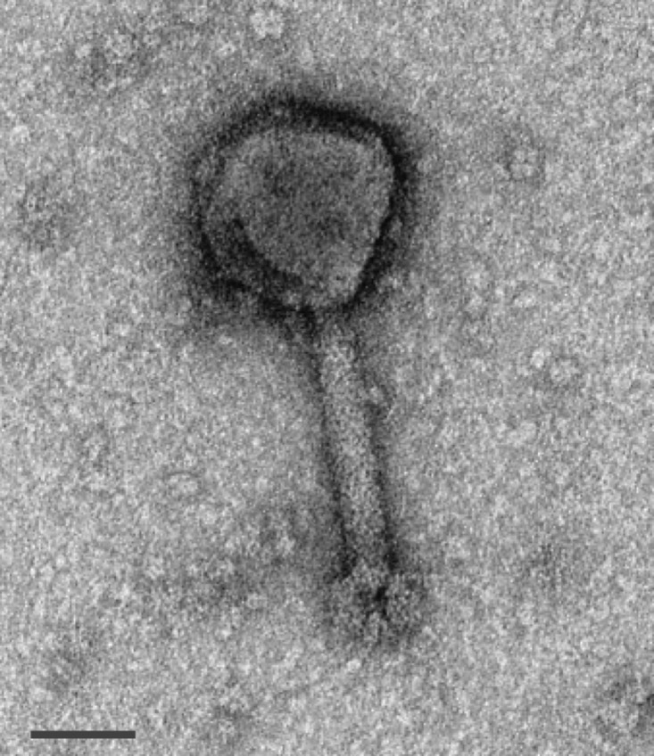
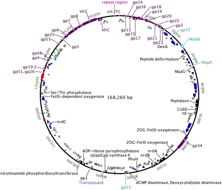
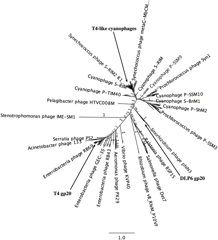
Increasing isolation of the extremely antibiotic resistant bacterium Stenotrophomonas maltophilia has caused alarm worldwide due to the limited treatment options available. A potential treatment option for fighting this bacterium is 'phage therapy', the clinical application of bacteriophages to selectively kill bacteria. Bacteriophage DLP6 (vB_SmoM-DLP6) was isolated from a soil sample using clinical isolate S. maltophilia strain D1571 as host. Host range analysis of phage DLP6 against 27 clinical S. maltophilia isolates shows successful infection and lysis in 13 of the 27 isolates tested. Transmission electron microscopy of DLP6 indicates that it is a member of the Myoviridae family. Complete genome sequencing and analysis of DLP6 reveals its richly recombined evolutionary history, featuring a core of both T4-like and cyanophage genes, which suggests that it is a member of the T4-superfamily. Unlike other T4-superfamily phages however, DLP6 features a transposase and ends with 229 bp direct terminal repeats. The isolation of this bacteriophage is an exciting discovery due to the divergent nature of DLP6 in relation to the T4-superfamily of phages.


Conflict of interest statement

Competing Interests: The authors have declared that no competing interests exist.

Figures

Fig 1
Fig 1. DLP6 phage morphology.
Liquid phage lysate was incubated on a carbon coated copper grid, stained with 4% uranyl acetate and visualized at 180,000-fold magnification by a transmission electron microscope. Scale bar represent 50 nm. The average capsid height measurement for DLP6 was 99 nm, average tail length of 144 nm and average tail width of 23 nm.
Fig 2
Fig 2. Genomic map of DLP6.
The scale (in bp) is shown in the outermost periphery of the genome along with late viral promoters (dark green) and terminators (dark red), as predicted by the algorithims in the software programs PHIRE and ARNold, respectively. Assigned functions for each predicted open reading frame are as follows: auxiliary metabolism (black), lysis (red), gene expression (light blue), phage morphogenesis (dark purple), DNA replication/repair (lilac), tRNA (bright green), repeat region (pink) and hypothetical (grey). Due to space constraints, genes are located either inside of or outside the circle thereby reducing overlap. The bp numbering of the circular map takes into account annealing of the direct repeats, reducing the genome length from 168,489 bp (- 229 bp) to 168,260 bp.
Fig 3
Fig 3. Predicted promoter sequence in DLP6.
Putative phage promoter consensus sequence identified using PHIRE [39] and plotted using WebLogo 3 [40].
Fig 4
Fig 4. Unrooted gp20 (portal vertex protein: AAY80_011) tree.
FastTree was used to generate the tree from a MUSCLE alignment between DLP6 gp20 and the top 250 BLASTP sequences. The local support value for each branch is shown on the tree and the bar is a marker of branch distance length. The clades featuring gp20 of T4 and gp20 of T4-superfamily cyanophages are indicated.
Fig 5
Fig 5. Circos plot of DLP6 and ΦM12 PROmer comparisons.
Green ribbons indicate regions of similarity between the two genomes at the protein level encoded on the same strand, and representing a similarity of greater than 32%, with an average similarity of 56%. Red ribbons indicate regions of similarity at the protein level encoded on opposite strands, representing a similarity of greater than 32%, with an average similarity of 56%. The scale (in kbp) is shown on the periphery of the plots. PROmer parameters: breaklen = 60, maxgap = 30, mincluster = 10, minmatch = 3.

Similar articles

Cited by

References

    1. World Health Organization. Antimicrobial resistance: global report on surveillance. World Health Organization; Geneva, Switzerland: 2014.
    1. Olaitan AO, Morand S, Rolain J-M. Emergence of colistin-resistant bacteria in humans without colistin usage: a new worry and cause for vigilance. Int. J. Antimicrob. Agents. 2016;47:1–3. 10.1016/j.ijantimicag.2015.11.009 - DOI - PubMed
    1. Brooke JS. Stenotrophomonas maltophilia: an emerging global opportunistic pathogen. Clin Microbiol Rev. 2012;25:2–41. 10.1128/CMR.00019-11 - DOI - PMC - PubMed
    1. Waters V, Atenafu EG, Lu A, Yau Y, Tullis E, Ratjen F. Chronic Stenotrophomonas maltophilia infection and mortality or lung transplantation in cystic fibrosis patients. J Cyst Fibros. 2013;12(5):482–6. 10.1016/j.jcf.2012.12.006 - DOI - PubMed
    1. Wainwright CE, France MW, O'Rourke P, Anuj S, Kidd TJ, Nissen MD, et al. Cough-generated aerosols of Pseudomonas aeruginosa and other Gram-negative bacteria from patients with cystic fibrosis. Thorax. 2009;64(11):926–31. 10.1136/thx.2008.112466 - DOI - PMC - PubMed

MeSH terms

LinkOut - more resources