. 2017 Mar 14;12(3):e0173823.
doi: 10.1371/journal.pone.0173823. eCollection 2017.

Adipocyte arrestin domain-containing 3 protein (Arrdc3) regulates uncoupling protein 1 (Ucp1) expression in white adipose independently of canonical changes in β-adrenergic receptor signaling

Affiliations

Adipocyte arrestin domain-containing 3 protein (Arrdc3) regulates uncoupling protein 1 (Ucp1) expression in white adipose independently of canonical changes in β-adrenergic receptor signaling

Shannon H Carroll et al. PLoS One. .

Erratum in

Abstract

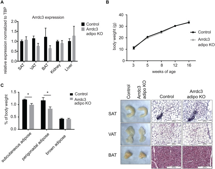
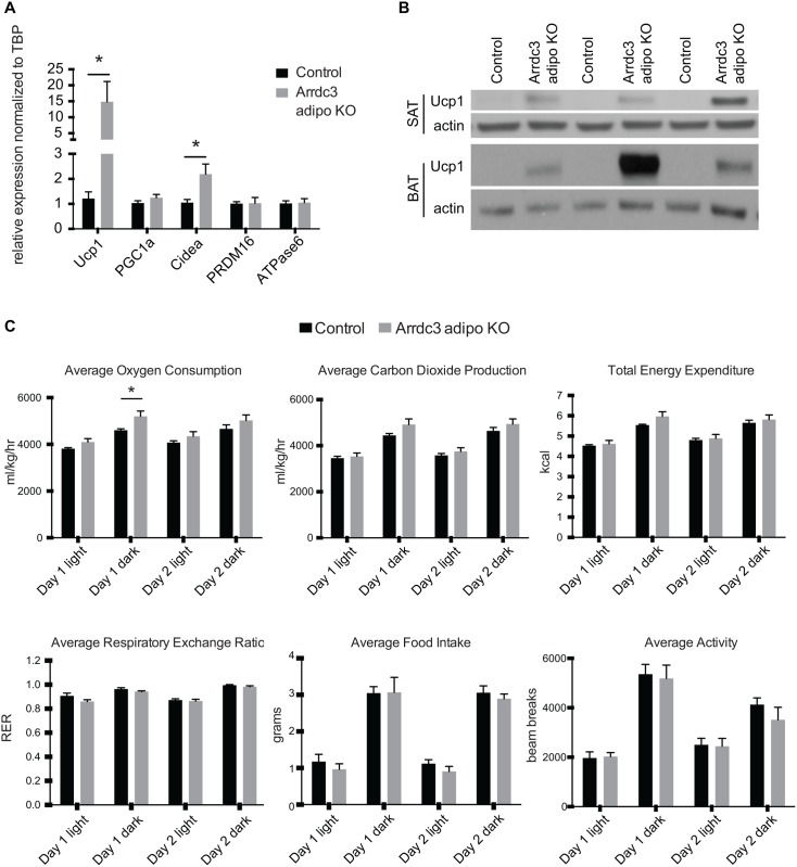
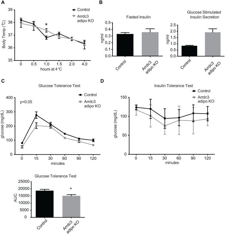
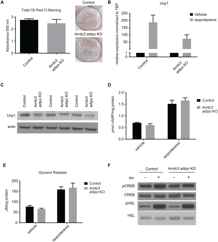
Adaptive thermogenesis and cold-induced activation of uncoupling protein 1 (Ucp1) in brown adipose tissue in rodents is well-described and attributed to sympathetic activation of β-adrenergic signaling. The arrestin domain containing protein Arrdc3 is a regulator of obesity in mice and also appears linked to obesity in humans. We generated a mouse with conditional deletion of Arrdc3, and here we present evidence that genetic ablation of Arrdc3 specifically in adipocytes results in increased Ucp1 expression in subcutaneous and parametrial adipose tissue. Although this increase in expression did not correspond with significant changes in body weight or energy expenditure, adipocyte-specific Arrdc3-null mice had improved glucose tolerance. It was previously hypothesized that Arrdc3 ablation leads to increased β-adrenergic receptor sensitivity; however, in vitro experiments show that Arrdc3-null adipocytes responded to β-adrenergic receptor agonist with decreased Ucp1 levels. Additionally, canonical β-adrenergic receptor signaling was not different in Arrdc3-null adipocytes. These data reveal a role for Arrdc3 in the regulation of Ucp1 expression in adipocytes. However, this adipocyte effect is insufficient to generate the obesity-resistant phenotype of mice with ubiquitous deletion of Arrdc3, indicating a likely role for Arrdc3 in cells other than adipocytes.


Conflict of interest statement

Competing Interests: The authors have declared that no competing interests exist.

Figures

Fig 1
Fig 1. Characterization of adipocyte-specific Arrdc3-null mice.
(A) To confirm adipocyte-specific deletion, Arrdc3 expression was measured in various tissues of Cre–(control) and Cre+ (Arrdc3-null) mice by quantitative PCR. Brown (BAT), parametrial (VAT) and subcutaneous adipose tissue (SAT) had significantly decreased Arrdc3 expression while there was no significant difference in liver or kidney (n = 3–4). (B) Adipocyte-specific Arrdc3-null mice and littermate controls were weighed for 16 weeks and no differences in body weight were found (n = 4–10). (C) Specific adipose depots of female mice were weighed and normalized to total body weight. Subcutaneous (SAT) and parametrial (VAT) adipose tissue from adipocyte-specific Arrdc3-null mice weighed significantly less than controls (n = 5). (D) Representative macroscopic (formaldehyde fixed tissue) and microscopic appearance of subcutaneous (SAT), parametrial (VAT) and brown (BAT) adipose tissue from adipocyte-specific Arrdc3-null and control mice. Paraffin tissue sections were stained with hematoxylin and eosin and images were taken at 40x.
Fig 2
Fig 2. Increased expression of Ucp1 in white adipose tissue of adipocyte-specific Arrdc3-null mice.
(A) Quantitative PCR analysis of gene expression in subcutaneous adipose tissue (n = 5–9). (B) Western analysis of Ucp1 protein expression in subcutaneous (SAT) and parametrial (VAT) adipose tissue. (C) 48 hours of CLAMS analysis of adipocyte-specific Arrdc3-null and control mice at 28°C ambient temperature (n = 5). *p≤ 0.05.
Fig 3
Fig 3. Effect of adipocyte-specific deletion of Arrdc3 on body temperature and glucose homeostasis.
(A) Control and adipocyte-specific Arrdc3-null mice were exposed to 4°C for four hours and core body temperature was measured at indicated time points (n = 4). (B) Blood insulin levels of control and adipocyte-specific Arrdc3-null mice after an overnight fast and after 2 minutes of i.p. glucose administration (n = 3). (C) Glucose tolerance testing of control and adipocyte-specific Arrdc3-null mice (n = 3). (D) Insulin tolerance testing of control and adipocyte-specific Arrdc3-null mice (n = 3). *p<0.05.
Fig 4
Fig 4. β-adrenergic signaling in Arrdc3-null adipocytes in vitro.
(A) Quantification of total Oil Red O staining of control and adipocyte-specific Arrdc3-null stromal vascular fractions after adipogenic treatment (n = 4 mice/group). Representative Oil Red O staining of stromal vascular fraction-derived adipocytes from control and adipocyte-specific Arrdc3-null mice. (B) Quantitative PCR analysis of Ucp1 upregulation upon 4 hours of isoproterenol treatment of control and Arrdc3-null cells (n = 4 mice/group). (C) Western analysis of Ucp1 protein expression upon 24 hours of isoproterenol treatment in control and Arrdc3-null cells. (D) cAMP concentration in control and Arrdc3-null cells upon 5 minutes of isoproterenol treatment (n = 3 mice/group). (E) Glycerol concentrations of control and Arrdc3-null cell media after 3 hours of isoproterenol treatment (n = 4 mice/group). (F) Western analysis of CREB and HSL phosphorylation of control and Arrdc3-null cells upon 5 minutes of isoproterenol treatment (n = 3 mice/group).
Fig 5
Fig 5. Arrdc3 deletion increases PPAR target gene expression in adipocytes.
Quantitative PCR array analysis of gene expression of PPAR target genes, PPAR cofactors and PPARs and associated transcription factors in control versus Arrdc3 SVF-derived adipocytes in vitro (n = 3 mice per group). Only significantly different genes are displayed. *p≤ 0.05.

Similar articles

Cited by

References

    1. Enerback S, Jacobsson A, Simpson EM, Guerra C, Yamashita H, et al. (1997) Mice lacking mitochondrial uncoupling protein are cold-sensitive but not obese. Nature 387: 90–94. - PubMed
    1. Cohen P, Spiegelman BM (2015) Brown and Beige Fat: Molecular Parts of a Thermogenic Machine. Diabetes 64: 2346–2351. 10.2337/db15-0318 - DOI - PMC - PubMed
    1. Keipert S, Jastroch M (2014) Brite/beige fat and UCP1—is it thermogenesis? Biochim Biophys Acta 1837: 1075–1082. 10.1016/j.bbabio.2014.02.008 - DOI - PubMed
    1. Kim SH, Plutzky J (2016) Brown Fat and Browning for the Treatment of Obesity and Related Metabolic Disorders. Diabetes Metab J 40: 12–21. 10.4093/dmj.2016.40.1.12 - DOI - PMC - PubMed
    1. Azzu V, Jastroch M, Divakaruni AS, Brand MD (2010) The regulation and turnover of mitochondrial uncoupling proteins. Biochim Biophys Acta 1797: 785–791. 10.1016/j.bbabio.2010.02.035 - DOI - PMC - PubMed