

[Cervicothoracic cystic lymphangioma: about a case]
- PMID: 28292151
- PMCID: PMC5326052
- DOI: 10.11604/pamj.2016.25.189.9363
[Cervicothoracic cystic lymphangioma: about a case]
Abstract
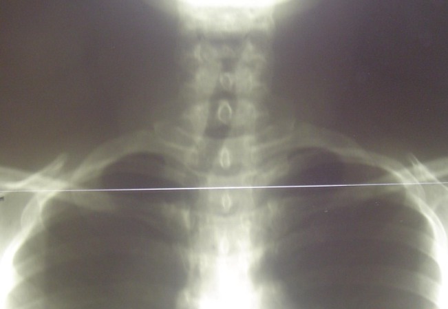
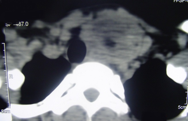
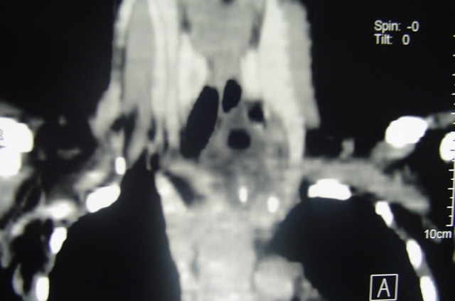
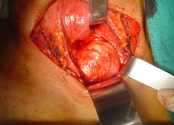
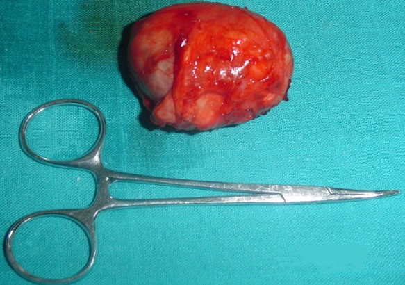
Cervicothoracic cystic lymphangiomas are rare benign tumors developing from sequestration of embryonic lymph sac which is gradually filled with lymph fluid. The diagnosis is based on clinical signs (laterocervical swelling) and imaging (ultrasound and CT scan), then confirmed by histology after surgery which constitutes the basis of treatment. We here report a case of cervicothoracic cystic lymphangioma and review of the literature.
Les lymphangiomes kystiques cervico-thoraciques sont des tumeurs bénignes rares, ils proviendraient d'une séquestration du sac lymphatique embryonnaire qui se remplirait progressivement de liquide lymphatique. Le diagnostic est évoqué par la clinique (tuméfaction latéro-cervicale) et l'imagerie (échographie et tomodensitométrie), puis confirmé par l'histologie après la chirurgie qui constitue la base du traitement. Nous rapportons un cas de lymphangiome kystique cervico-thoracique avec une revue de la littérature.
Keywords: Cystic lymphangioma; chest; neck.
Conflict of interest statement
Les auteurs ne déclarent aucun conflit d'intérêt.
Figures
References
-
- Handa R, Kale R, Upadhyay KK. Isolated médiastinal lymphangioma herniating through the intercostal space. Asian J Surg. 2004;27(3):241–242. - PubMed
-
- N'dri K, Adjenou V, Konan A, Gbazi GC, Mensah GD, Aguehounde C, Abby BC. Lymphangiome kystique cervical: apport de l'échographie et de la tomodensitométrie a propos d'un cas. Médecine d'Afrique noire. 1996;43(4):237–239.
-
- Haenggeli A, Becker M, Crescentino V, Kurt AM, Lemann W. Lymphangiome kystique cervical et lymphangiomatoses : Présentation d'un cas. Médecine et hygiène. 1997;55(2183):2088–2090.
-
- Poyraz AS, Kilic D, Hatipoglu A, Ozulku M, Sar A, Bilezikci B. Cystic lymphangioma confined to mediastinum in adult. Jpn J Thorac Cardiovasc Surg. 2004;52(12):567–569. - PubMed
-
- Özlem E, Özçakar L, Inanici F. Lymphangiome kystique du muscle quadriceps: un diagnostic rassurant pour une douleur du genou. Revue de Rhumatisme. 2005;72(5):440–442.
Publication types
MeSH terms
LinkOut - more resources
Full Text Sources
Other Literature Sources
