. 2017 Mar 15;14(1):47.
doi: 10.1186/s12974-017-0819-4.

Microglial-derived microparticles mediate neuroinflammation after traumatic brain injury

Affiliations

Microglial-derived microparticles mediate neuroinflammation after traumatic brain injury

Alok Kumar et al. J Neuroinflammation. .

Abstract

Background: Local and systemic inflammatory responses are initiated early after traumatic brain injury (TBI), and may play a key role in the secondary injury processes resulting in neuronal loss and neurological deficits. However, the mechanisms responsible for the rapid expansion of neuroinflammation and its long-term progression have yet to be elucidated. Here, we investigate the role of microparticles (MP), a member of the extracellular vesicle family, in the exchange of pro-inflammatory molecules between brain immune cells, as well as their transfer to the systemic circulation, as key pathways of inflammation propagation following brain trauma.

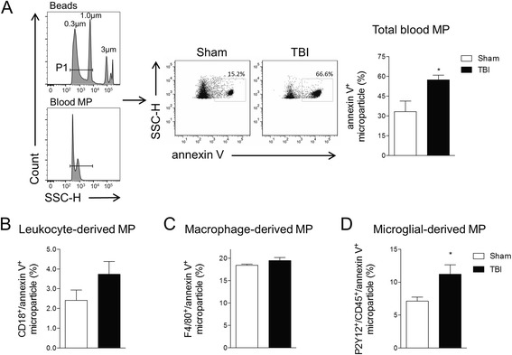
Methods: Adult male C57BL/6 mice were subjected to controlled cortical impact TBI for 24 h, and enriched MP were isolated in the blood, while neuroinflammation was assessed in the TBI cortex. MP were characterized by flow cytometry, and MP content was assayed using gene and protein markers for pro-inflammatory mediators. Enriched MP co-cultured with BV2 or primary microglial cells were used for immune propagation assays. Enriched MP from BV2 microglia or CD11b-positive microglia from the TBI brain were stereotactically injected into the cortex of uninjured mice to evaluate MP-related seeding of neuroinflammation in vivo.

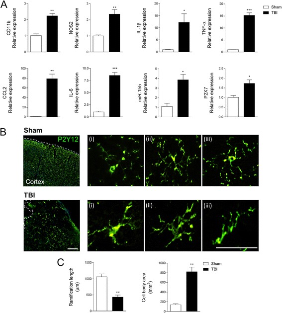
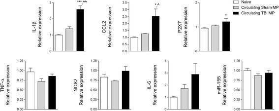
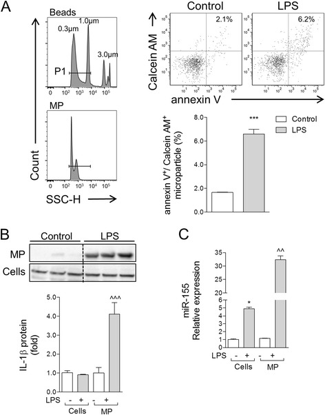
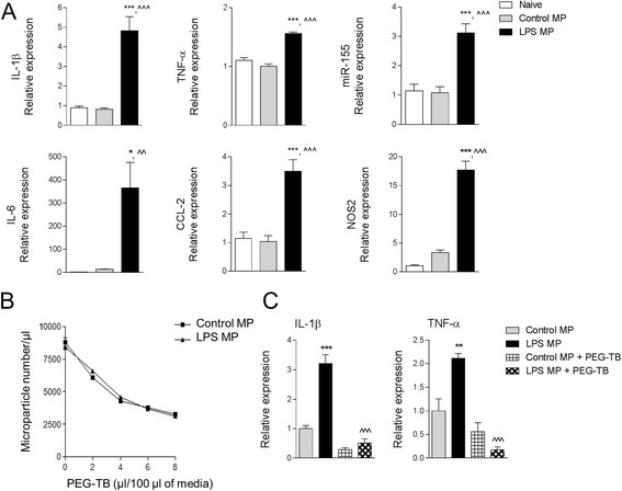
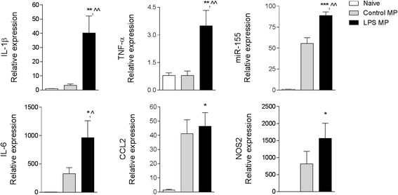
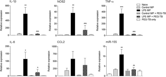
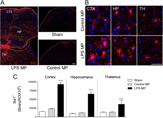
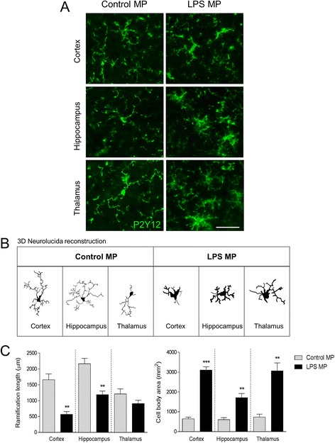
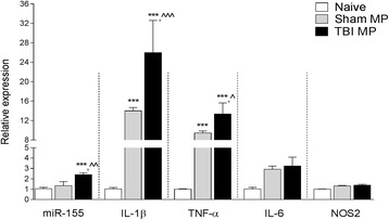
Results: As the neuroinflammatory response is developing in the brain after TBI, microglial-derived MP are released into the circulation. Circulating enriched MP from the TBI animals can activate microglia in vitro. Lipopolysaccharide stimulation increases MP release from microglia in vitro and enhances their content of pro-inflammatory mediators, interleukin-1β and microRNA-155. Enriched MP from activated microglia in vitro or CD11b-isolated microglia/macrophage from the TBI brain ex vivo are sufficient to initiate neuroinflammation following their injection into the cortex of naïve (uninjured) animals.

Conclusions: These data provide further insights into the mechanisms underlying the development and dissemination of neuroinflammation after TBI. MP loaded with pro-inflammatory molecules initially released by microglia following trauma can activate additional microglia that may contribute to progressive neuroinflammatory response in the injured brain, as well as stimulate systemic immune responses. Due to their ability to independently initiate inflammatory responses, MP derived from activated microglia may provide a potential therapeutic target for other neurological disorders in which neuroinflammation may be a contributing factor.

Keywords: Interleukin-1β; Microglia; Microparticles; Neuroinflammation; Traumatic brain injury; miR-155.


Figures

Fig. 1
Fig. 1
Microglial-derived MP are increased in the blood following TBI. Flow cytometry analysis of enriched MP in the blood from sham and TBI mice at 24 h post-injury. a Representation of gating strategy used to characterize MP using SSC-H and standard microbeads (300- to 1000-nm diameter). Standard microbeads (P1 gated population) were used as an internal control to determine the size of MP in the blood, and annexin V staining confirmed MP characteristics. At 24 h post-injury, total blood MP is increased in TBI mice compared with sham-injured mice. b Measurements of leukocyte-derived (CD18), macrophage-derived (F4/80), and microglial-derived (P2Y12/CD45) MP in the blood from sham and TBI mice at 24 h post-injury. Microglial-derived MP are significantly increased in TBI mice when compared with sham-injured mice (*p < 0.5 vs sham; Student’s t test; n = 6/group). Bars represent mean ± standard error of the mean (S.E.M.). Data represent results of three independent experiments
Fig. 2
Fig. 2
Microglial activation in the TBI brain at 24 h post-injury. a Gene expression analysis of microglia activation in the cortex of sham and TBI mice at 24 h post-injury. Microglial receptors (CD11b, P2X7) and pro-inflammatory mediators (NOS2, IL-1β, TNF-α, CCL2, IL-6, and miR-155) were significantly increased in the injured cortex at 24 h post-injury (*p < 0.05, **p < 0.01, and ***p < 0.001 vs sham-injured; Student’s t test; n = 6/group). b Immunofluorescence imaging for P2Y12-positive microglia in the cortex of sham and TBI mice at 24 h post-injury. Following TBI P2Y12-positive microglia transformed from a ramified morphology in sham to activated morphology displaying enlarged cell body, and thicker, and shorter, projections. Representative images taken at −2.06 mm from the bregma. Scale bar = 50 μm. c Morphological analysis of P2Y12-positive microglia using 3D-reconstruction Neurolucida software. When compared to sham-injured controls, P2Y12-positive microglia in the TBI cortex had reduced ramification length (**p < 0.01; Student’s t test) and an enlarged cell body area (**p < 0.01; Student’s t test; n = 6/group). Bars represent mean ± S.E.M.
Fig. 3
Fig. 3
Circulating blood MP released after TBI activate naive BV2 microglia. Enriched MP were isolated from the blood of sham-injured and TBI mice and were co-cultured with naïve BV2 microglia cells for 24 h. Microglial receptors (P2X7) and pro-inflammatory mediators (IL-1β and CCL2) were significantly increased in BV2 microglia treated with circulating TBI MP (*p < 0.05 and ***p < 0.001 vs naïve; ^p < 0.05 and ^^p < 0.01 vs sham MP; one-way ANOVA with Student-Newman-Keuls correction for multiple comparisons; n = 6/group). There were no significant differences in TNF-α, NOS2, IL-6, and miR-155 expression between treatment groups. Bars represent mean ± S.E.M.
Fig. 4
Fig. 4
Pro-inflammatory mediators are enriched in MP following lipopolysaccharide stimulation of BV2 microglia. a Calcein AM-stained BV2 microglia were stimulated with LPS (20 ng/ml) for 24 h, and MP were isolated by differential centrifugation and stained with anti-annexin V, prior to characterization by flow cytometry. When compared to MP levels in control BV2 microglia, LPS stimulation significantly increased the number of annexin V/calcein AM-positive MP (***p < 0.001 vs control; Student’s t test; n = 4/group). b IL-1β protein expression in enriched MP vs cell lysates of control and LPS-stimulated BV2 microglia. Western blot analysis demonstrated that IL-1β protein was significantly increased in enriched MP following LPS stimulation (^^^p < 0.001 vs control MP; one-way ANOVA with Student-Newman-Keuls correction for multiple comparisons; n = 3/group). Lanes 1, 2, and 3 in both control and LPS refer to sample replicates. c miR-155 expression in enriched MP vs cell lysates of control and LPS-stimulated BV2 microglia. miR-155 was significantly increased in cell lysates of BV2 microglia following LPS stimulation (*p < 0.05 vs control cells), and its expression was elevated further in enriched MP (^^p < 0.01 vs control MP; one-way ANOVA with Student-Newman-Keuls correction for multiple comparisons; n = 4/group). Bars represent mean ± S.E.M. Data represent results of three independent experiments
Fig. 5
Fig. 5
Lipopolysaccharide-stimulated MP activate BV2 microglia. a Enriched MP were isolated from control and LPS-stimulated BV2 microglia and were co-cultured with naïve BV2 microglia for 24 h. Pro-inflammatory mediators (IL-1β, TNF-α, miR-155, IL-6, CCL2, and NOS2) were significantly increased in BV2 microglia treated with LPS MP (*p < 0.05 and ***p < 0.001 vs naïve; ^^p < 0.01 and ^^^p < 0.001 vs control MP; one-way ANOVA with Student-Newman-Keuls correction for multiple comparisons; n = 4/group). Data represent results of three independent experiments. b MP neutralization using PEG-TB. Enriched MP from control and LPS-stimulated BV2 microglia were incubated with increasing concentrations of PEG-TB for 1 h, and number of MP were quantified by flow cytometry. 6 μl PEG-TB/100 μl resulted in significant depletion of MP under both conditions. c Naïve BV2 microglia were co-cultured with control or LPS-stimulated MP ± PEG-TB (6 μl/100 μl) for 24 h. LPS MP treatment increased IL-1β and TNF-α in BV2 microglia (**p < 0.01 and ***p < 0.001 vs control MP), whereas co-treatment with PEG-TBI resulted in a significant decrease in IL-1β and TNF-α expression (^^^p < 0.001 vs LPS MP; one-way ANOVA with Student-Newman-Keuls correction for multiple comparisons; n = 6/group). Bars represent mean ± S.E.M.
Fig. 6
Fig. 6
Lipopolysaccharide-stimulated MP activate primary cortical microglia. Enriched MP were isolated from control and LPS-stimulated BV2 microglia and were co-cultured with primary cortical microglia for 24 h. Pro-inflammatory mediators (IL-1β, TNF-α, miR-155, IL-6, CCL2, and NOS2) were significantly increased in primary microglia treated with LPS MP (*p < 0.05, **p < 0.001, and ***p < 0.001 vs naïve; ^p < 0.05 and ^^p < 0.01 vs control MP; one-way ANOVA with Student-Newman-Keuls correction for multiple comparisons; n = 5/group). Bars represent mean ± S.E.M.
Fig. 7
Fig. 7
Lipopolysaccharide-stimulated MP increase neuroinflammation in the cortex of uninjured mice. Enriched MP were isolated from control and LPS-stimulated BV2 microglia and were treated ± PEG-TB (6 μl/100 μl) prior to being stereotactically injected into the cortex of adult male C57BL/6 mice. Markers of cortical neuroinflammation were measured at 24 h postinjection. There was a significant increase in pro-inflammatory mediators (IL-1β, TNF-α, miR-155, IL-6, and NOS2) in the cortex of LPS MP-injected mice (**p < 0.01, ***p < 0.001 vs control MP-injected group). Neutralization of LPS MP prior to injection resulted in a significant decrease in each pro-inflammatory mediator (^p < 0.05, ^^p < 0.01, ^^^p < 0.001 vs LPS MP-injected group; one-way ANOVA with Student-Newman-Keuls correction for multiple comparisons; n = 6/group). Bars represent mean ± S.E.M.
Fig. 8
Fig. 8
Lipopolysaccharide-stimulated MP increase microglial activation in the cortex of uninjured mice. Enriched MP were isolated from control and LPS-stimulated BV2 microglia and were stereotactically injected into the cortex of adult male C57BL/6 mice. Iba-1 immunocytochemistry was performed at 7 days postinjection. a Representative Iba-1 staining (red) in the cortex (CTX), hippocampus (HP), and thalamus (TH). Images taken at −2.06 mm from the bregma; scale bar = 50 μm. b High-magnification images in control MP- and LPS MP-injected mice in the CTX, HP, and TH. LPS MP-injected Iba-1-positive microglia had enlarged cell body and thicker projection indicative of increased activation status. Scale bar = 100 μm. c Quantification of Iba-1 staining in the cortex, hippocampus, and thalamus at 7 days postinjection. There was a significant increase in Iba-1 immunoreactivity in the LPS MP-injected group when compared to the control MP-treated group (***p < 0.001 vs control MP; one-way ANOVA with Student-Newman-Keuls correction for multiple comparisons; n = 4/group). Bars represent mean ± S.E.M.
Fig. 9
Fig. 9
Lipopolysaccharide-stimulated MP alter P2Y12 microglial morphology of the cortex of uninjured mice. Enriched MP were isolated from control and LPS-stimulated BV2 microglia and were stereotactically injected into the cortex of adult male C57BL/6 mice. P2Y12 immunocytochemistry was performed at 7 days postinjection. a High-magnification images of P2Y12-positive microglia (green) in control MP- and LPS MP-injected mice in the cortex (CTX), hippocampus (HP), and thalamus (TH). LPS MP-injected P2Y12-positive microglia have enlarged cell body and thicker projection indicative of increased activation status. Scale bar = 100 μm. b P2Y12-positive microglia in the CTX, HP, and TH of control MP- and LPS MP-injected mice. c Morphological analysis of P2Y12-positive microglia using 3D-reconstruction Neurolucida software. When compared to the control MP-injected group, P2Y12-positive microglia in the LPS MP-injected group had reduced ramification length in the cortex and hippocampus (**p < 0.01; Student’s t test), but not in the thalamus. In addition, the LPS MP-injected group had enlarged cell body area in each region (**p < 0.01 and ***p < 0.001 vs control MP; Student’s t test; n = 4/group). Bars represent mean ± S.E.M.
Fig. 10
Fig. 10
MP isolated from CD11b-isolated microglia/macrophages following TBI increase neuroinflammation in the cortex of uninjured mice. CD11b-microglia/macrophages in the cortex of sham and TBI were isolated at 7 days post-injury and cultured for 24 h prior to collecting MP. Enriched MP were stereotactically injected into the cortex of adult male C57BL/6 mice and markers of cortical neuroinflammation were measured at 24 h postinjection. There was a significant increase in pro-inflammatory mediators (IL-1β and TNF-α) in the cortex of the control MP-injected group (***p < 0.001 vs naïve group). When compared to the control MP-injected group, there was a further significant increase in pro-inflammatory mediators (IL-1β, TNF-α, and miR-155) in the cortex of the TBI MP-injected group (^p < 0.05, ^^p < 0.01, ^^^p < 0.001 vs control MP-injected group; one-way ANOVA with Student-Newman-Keuls correction for multiple comparisons; n = 5/group). Bars represent mean ± S.E.M.

Similar articles

Cited by

References

    1. Burnier L, Fontana P, Kwak BR, Angelillo-Scherrer A. Cell-derived microparticles in haemostasis and vascular medicine. Thromb Haemost. 2009;101:439–451. - PubMed
    1. Bohman LE, Riley J, Milovanova TN, Sanborn MR, Thom SR, Armstead WM. Microparticles impair hypotensive cerebrovasodilation and cause hippocampal neuronal cell injury after traumatic brain injury. J Neurotrauma. 2016;33:168–174. doi: 10.1089/neu.2015.3885. - DOI - PMC - PubMed
    1. Davizon P, Munday AD, Lopez JA. Tissue factor, lipid rafts, and microparticles. Semin Thromb Hemost. 2010;36:857–864. doi: 10.1055/s-0030-1267039. - DOI - PubMed
    1. Zappulli V, Friis KP, Fitzpatrick Z, Maguire CA, Breakefield XO. Extracellular vesicles and intercellular communication within the nervous system. J Clin Invest. 2016;126:1198–1207. doi: 10.1172/JCI81134. - DOI - PMC - PubMed
    1. Castro-Seoane R, Hummerich H, Sweeting T, Tattum MH, Linehan JM, Fernandez de Marco M, Brandner S, Collinge J, Klohn PC. Plasmacytoid dendritic cells sequester high prion titres at early stages of prion infection. PLoS Pathog. 2012;8:e1002538. doi: 10.1371/journal.ppat.1002538. - DOI - PMC - PubMed

Publication types

MeSH terms