. 2017 Mar 14;5(1):22.
doi: 10.1186/s40478-017-0426-8.

Age-dependent dopamine transporter dysfunction and Serine129 phospho-α-synuclein overload in G2019S LRRK2 mice

Affiliations

Age-dependent dopamine transporter dysfunction and Serine129 phospho-α-synuclein overload in G2019S LRRK2 mice

Francesco Longo et al. Acta Neuropathol Commun. .

Abstract

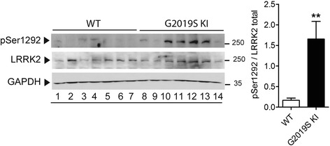
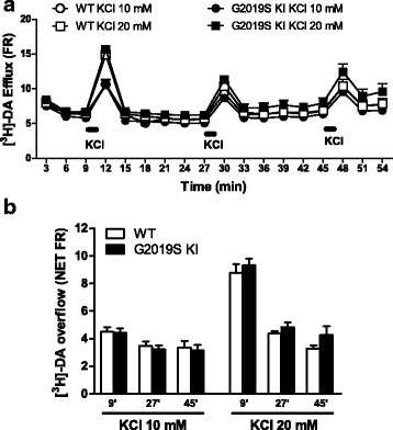
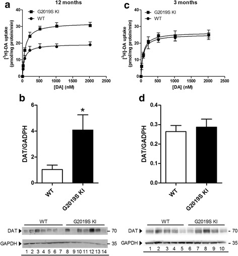
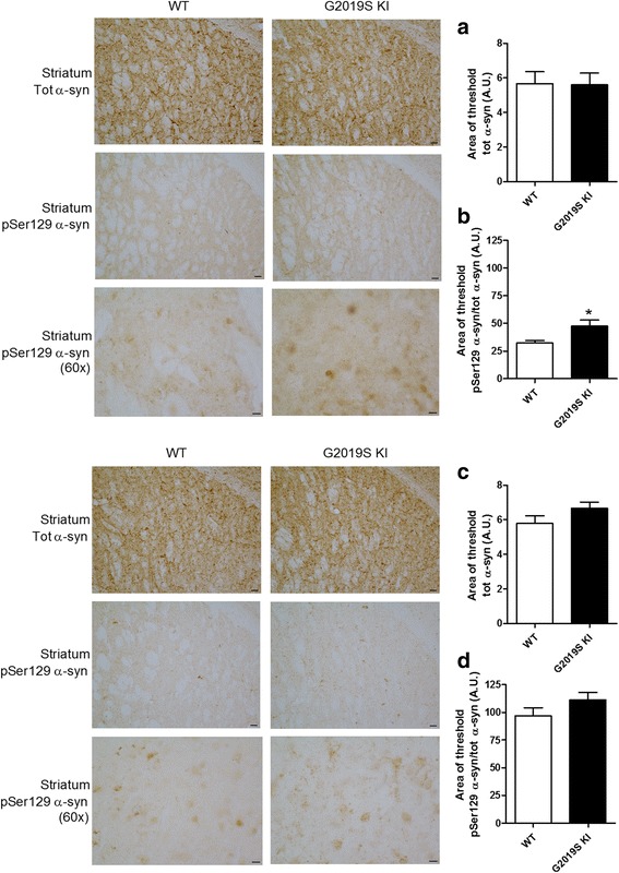
Mutations in the leucine-rich repeat kinase 2 (LRRK2) gene are the most common genetic cause of Parkinson's disease. Here, we investigated whether the G2019S LRRK2 mutation causes morphological and/or functional changes at nigro-striatal dopamine neurons. Density of striatal dopaminergic terminals, nigral cell counts, tyrosine hydroxylase protein levels as well as exocytotic dopamine release measured in striatal synaptosomes, or striatal extracellular dopamine levels monitored by in vivo microdialysis were similar between ≥12-month-old G2019S knock-in mice and wild-type controls. In vivo striatal dopamine release was insensitive to the LRRK2 inhibitor Nov-LRRK2-11, and was elevated by the membrane dopamine transporter blocker GBR-12783. However, G2019S knock-in mice showed a blunted neurochemical and motor activation response to GBR-12783 compared to wild-type controls. Western blot and dopamine uptake analysis revealed an increase in dopamine transporter levels and activity in the striatum of 12-month-old G2019S KI mice. This phenotype correlated with a reduction in vesicular monoamine transporter 2 levels and an enhancement of vesicular dopamine uptake, which was consistent with greater resistance to reserpine-induced hypolocomotion. These changes were not observed in 3-month-old mice. Finally, Western blot analysis revealed no genotype difference in striatal levels of endogenous α-synuclein or α-synuclein bound to DOPAL (a toxic metabolite of dopamine). However, Serine129-phosphorylated α-synuclein levels were higher in 12-month-old G2019S knock-in mice. Immunohistochemistry confirmed this finding, also showing no genotype difference in 3-month-old mice. We conclude that the G2019S mutation causes progressive dysfunctions of dopamine transporters, along with Serine129-phosphorylated α-synuclein overload, at striatal dopaminergic terminals, which are not associated with dopamine homeostasis dysregulation or neuron loss but might contribute to intrinsic dopaminergic terminal vulnerability. We propose G2019S knock-in mice as a presymptomatic Parkinson's disease model, useful to investigate the pathogenic interaction among genetics, aging, and internal or environmental factors leading to the disease.

Keywords: DAT; G2019S knock-in; LRRK2; Parkinson’s disease; VMAT2; α-synuclein.


Figures

Fig. 1
Fig. 1
Phosphorylation levels of LRRK2 at Ser1292 (pSer1292) are elevated in G2019S knock-in (KI) mice. Striatal pSer1292 and total LRRK2 levels were measured by Western blotting in 12-month-old G2019S KI mice and age-matched WT controls. Representative blots (left) and quantification (right) are shown. Data are expressed as pSer1292 LRRK2/total LRRK2 and are means ± SEM of 7 animals per group. Statistical analysis was performed with the Student t-test, two tailed for unpaired data. **p < 0.01, different from WT
Fig. 2
Fig. 2
The integrity of nigro-striatal dopaminergic neurons is preserved in G2019S knock-in (KI) mice. Stereological count of nigral DA neurons (a) and density of tyrosine hydroxylase (TH) positive striatal nerve terminals (b), with representative images, in 12-month-old G2019S KI mice and age-matched WT littermates. Western blotting analysis of striatal TH levels in 12-month-old G2019S KI mice and age-matched WT controls (c). Data are expressed as absolute values and are means ± SEM of 8 (a-b) and 4 (c) animals per group
Fig. 3
Fig. 3
Dopamine (DA) release is preserved in G2019S knock-in (KI) mice. [3H]-DA preloaded synaptosomes obtained from the striata of 12-month-old G2019S KI mice and age-matched WT littermates were continuously superfused with Krebs and stimulated with 3 pulses (90 s) of 10 mM or 20 mM K+ (18 min apart). DA release has been expressed as fractional release (FR; i.e. tritium efflux expressed as percentage of the tritium content in the filter at the onset of the corresponding collection period; a), or NET FR (i.e. K+-evoked tritium overflow as percent of the tritium content in the filter at the onset of the corresponding collection period; b). Data are means ± SEM of 9 determinations per group
Fig. 4
Fig. 4
Acute blockade of LRRK2 kinase activity does not affect striatal dopamine (DA) levels whereas acute DAT blockade evoked blunted neurochemical and behavioral responses in G2019S knock-in (KI) mice in vivo. Microdialysis was performed in the dorsolateral striatum of 19-month-old G2019S KI mice and with age-matched wild-type (WT) littermates (WT) (a, b). Mice were then challenged with the LRRK2 kinase inhibitor Nov-LRRK2-11 (10 mg/kg, i.p.) or the DAT blocker GBR-12783 (20 mg/kg, i.p.). Dialysate levels of DA are expressed as absolute values (nM) and are mean ± SEM of 5 WT and 6 G2019S KI mice (a), or 6 WT and 9 G2019S KI mice (b). Motor responses of 12-month-old mice to GBR-12783 (6 mg/kg, i.p.) or saline administration (c-e). Motor activity was assessed using the bar (c), drag (d) and rotarod (e) tests, before (baseline) and after (20 and 90 min) drug administration, and was expressed as percentage of performance at baseline. Data are means ± SEM of 12–17 (WT) or 13–17 (G2019S KI) mice per group. Statistical analysis was performed using one-way RM ANOVA (a-b) or conventional ANOVA (c-e) followed by the Newman–Keuls test for multiple comparisons. *p < 0.05, ** p < 0.01 significantly different from baseline values; # p < 0.05, ## p < 0.01 significantly different from saline
Fig. 5
Fig. 5
Age-dependent dysfunction of DAT expression and function in G2019S knock-in (KI) mice. Kinetic analysis of [3H]-DA uptake in synaptosomes (a, c), and Western blotting analysis of DAT protein levels (and representative blots) (b, d) were performed in the striata of 12-month-old (a, b) and 3-month-old (c, d) G2019S KI mice in comparison with age-matched WT controls. Values are expressed as mean ± SEM of n = 4 (uptake) or n = 3 (Western blotting) independent experiments performed in duplicate. Statistical analysis was performed using the Student t-test, two-tailed for unpaired data. *p < 0.05, different from WT
Fig. 6
Fig. 6
Age-dependent dysfunction of VMAT2 expression and function in G2019S knock-in (KI) mice. Motor activity in 12-month-old G2019S KI mice and wild-type (WT) littermates treated with reserpine (1 mg/kg, i.p.) or saline, and challenged in the bar (a), drag (b) and rotarod (c) tests, before (baseline) and after (24 and 48 h) drug administration. Motor performance was expressed as percentage of performance at baseline. Data are means ± SEM of n = 14–15 mice per group and were analyzed using conventional ANOVA followed by the Newman–Keuls test for multiple comparisons. # p < 0.05, ## p < 0.01 significantly different from saline. Kinetic analysis of [3H]-DA uptake in whole-brain vesicles and Western blotting of VMAT2 levels in the striata from 12-month-old (d-f) or 3-month old (g- i) G2019S KI mice and WT littermates. In Western blotting, two different anti-VMAT2 antibodies were used, one commercially available (e, h; Sigma) and another developed by Miller lab (f, i) (see Methods). Data are expressed as mean ± SEM of 4 mice (d- f), 3 mice (g) or 5 mice (h, i) per group, performed in duplicate. Statistical analysis was performed by the Student t-test, two-tailed for unpaired data. **p < 0.01, different from WT
Fig. 7
Fig. 7
DOPAL-modified α-synuclein (α-syn) levels are unchanged whereas Ser129-phosphorylated α-synuclein (pSer129 α-syn) levels are elevated in G2019K knock-in (KI) mice. Relative quantification and representative blots of DOPAL-bound α-syn pull-down with aminophenylboronic acid (APBA) resin of striata from 12-month-old G2019S KI mice and age-matched WT controls (a). In the same preparation, pSer129 α-syn levels (c) were quantified relatively to α-syn levels (b). Data are expressed as mean ± SEM of n = 11 mice per group. Statistical analysis was performed by the Student t-test, two tailed for unpaired data. **p < 0.01 different from WT
Fig. 8
Fig. 8
Age-dependent overload of Serine129-phosphorylated α-synuclein (pSer129 α-syn) in G2019S knock-in (KI) mice. Representative microphotographs and relative quantifications of α-syn and pSer129 α-syn immunostaining in the striatum of 12-month-old (a, b) and 3-month-old (c, d) G2019S KI mice and WT controls. Data are expressed as mean ± SEM of 8 (a, b) or 6 (c, d) mice per group. Statistical analysis was performed by the Student t-test, two tailed for unpaired data. *p < 0.05 different from WT

References

    1. Anderson JP, Walker DE, Goldstein JM, de Laat R, Banducci K, Caccavello RJ, Barbour R, Huang J, Kling K, Lee M, Diep L, Keim PS, Shen X, Chataway T, Schlossmacher MG, Seubert P, Schenk D, Sinha S, Gai WP, Chilcote TJ. Phosphorylation of Ser-129 is the dominant pathological modification of alpha-synuclein in familial and sporadic Lewy body disease. J Biol Chem. 2006;281:29739–29752. doi: 10.1074/jbc.M600933200. - DOI - PubMed
    1. Arcuri L, Viaro R, Bido S, Longo F, Calcagno M, Fernagut PO, Zaveri NT, Calo G, Bezard E, Morari M. Genetic and pharmacological evidence that endogenous nociceptin/orphanin FQ contributes to dopamine cell loss in Parkinson's disease. Neurobiol Dis. 2016;89:55–64. doi: 10.1016/j.nbd.2016.01.016. - DOI - PMC - PubMed
    1. Beccano-Kelly DA, Kuhlmann N, Tatarnikov I, Volta M, Munsie LN, Chou P, Cao LP, Han H, Tapia L, Farrer MJ, Milnerwood AJ. Synaptic function is modulated by LRRK2 and glutamate release is increased in cortical neurons of G2019S LRRK2 knock-in mice. Front Cell Neurosci. 2014;8:301. doi: 10.3389/fncel.2014.00301. - DOI - PMC - PubMed
    1. Beccano-Kelly DA, Volta M, Munsie LN, Paschall SA, Tatarnikov I, Co K, Chou P, Cao LP, Bergeron S, Mitchell E, Han H, Melrose HL, Tapia L, Raymond LA, Farrer MJ, Milnerwood AJ. LRRK2 overexpression alters glutamatergic presynaptic plasticity, striatal dopamine tone, postsynaptic signal transduction, motor activity and memory. Hum Mol Genet. 2015;24:1336–1349. doi: 10.1093/hmg/ddu543. - DOI - PubMed
    1. Bezard E. Neuroprotection for Parkinson's disease: a call for clinically driven experimental design. Lancet Neurol. 2003;2:393. doi: 10.1016/S1474-4422(03)00432-0. - DOI - PubMed

Publication types

MeSH terms

Substances

LinkOut - more resources