Review
. 2017 Mar 15;144(6):976-985.
doi: 10.1242/dev.143529.

Embryoids, organoids and gastruloids: new approaches to understanding embryogenesis

Affiliations
Review

Embryoids, organoids and gastruloids: new approaches to understanding embryogenesis

Mijo Simunovic et al. Development. .

Abstract

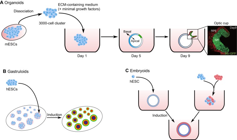
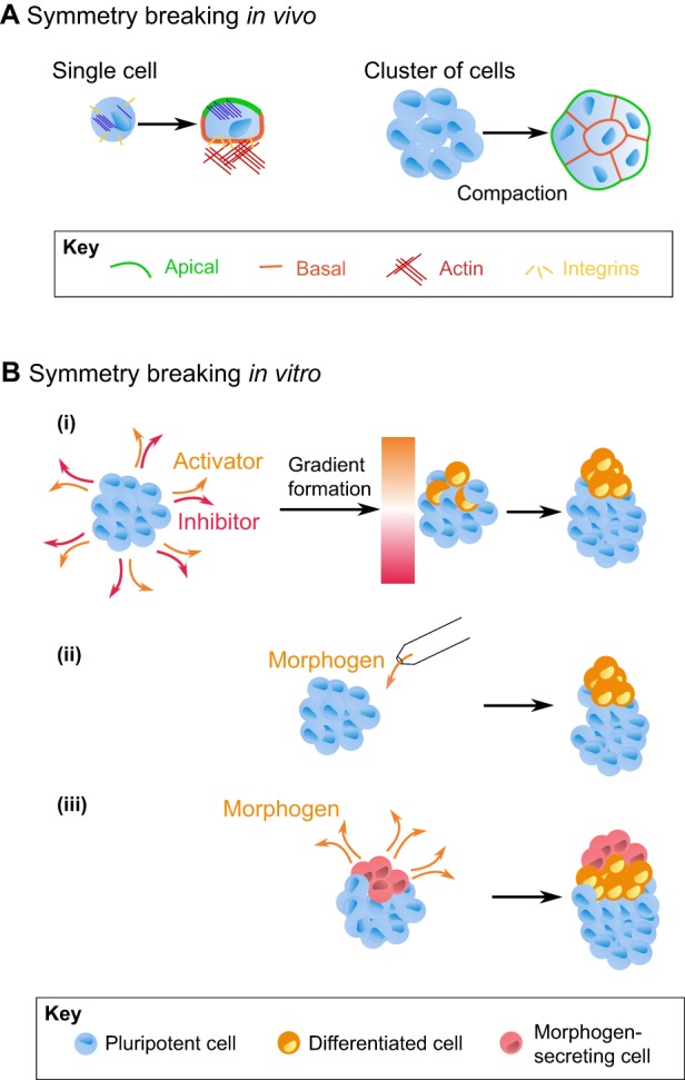
Cells have an intrinsic ability to self-assemble and self-organize into complex and functional tissues and organs. By taking advantage of this ability, embryoids, organoids and gastruloids have recently been generated in vitro, providing a unique opportunity to explore complex embryological events in a detailed and highly quantitative manner. Here, we examine how such approaches are being used to answer fundamental questions in embryology, such as how cells self-organize and assemble, how the embryo breaks symmetry, and what controls timing and size in development. We also highlight how further improvements to these exciting technologies, based on the development of quantitative platforms to precisely follow and measure subcellular and molecular events, are paving the way for a more complete understanding of the complex events that help build the human embryo.

Keywords: Early development; Patterning; Self-assembly; Self-organization; Symmetry breaking; Tissue mechanics.


Conflict of interest statement

Competing interests

The authors declare no competing or financial interests.

Figures

Fig. 1.
Fig. 1.
Self-organization into organoids, gastruloids and embryoids. (A) A cluster of dissociated mouse embryonic stem cells (mESCs) cultured in a medium containing extracellular matrix (ECM) proteins and minimal growth factors spontaneously self-organizes, first into a polarized quasi-spherical epithelial tissue, then later giving rise to a structure resembling an optic cup. Rx+ cells (green) mark the retinal anlage; Mitf+ cells (red) mark the epithelial shell of the optic cup. NR, neural retina; RPE, retinal pigment epithelium. Microscopy image adapted with permission (Eiraku et al., 2011). (B) Dissociated human embryonic stem cells (hESCs) are seeded on a surface patterned with polymerized ECM proteins, creating demarcated cell colonies of defined size and shape. Subsequent addition of morphogen may give rise to the patterned differentiation of cells. In the case of BMP4 induction, patterned cells form gastruloids with all germ layers (Deglincerti et al., 2016b; Warmflash et al., 2014). Colors in patterned cell colonies represent different germ layers. (C) Some routes by which cells can be induced to form a multilayered embryoid. Left pathway: hESCs form an organized 3D structure, and subsequent induction leads to pattern formation. Right pathway: the mixing of multiple cell types gives rise to sorting and differentiation into an organized embryoid.
Fig. 2.
Fig. 2.
Features of ESC self-organization and patterning. (A) A simple example of polarity-based self-organization. Dissociated ESCs on a surface coated with polymerized ECM proteins form a polarized epithelium, with cells exhibiting apical (green) and basolateral (orange) surfaces. (B) Sorting driven by the minimization of tissue surface energy. Cells with the strongest cell-cell interactions (based on adhesive energy or tissue tension) migrate toward the interior of a cell cluster, while those with the weakest cell-cell interactions migrate toward the exterior. (C) Lumen formation in ESCs. Cells embedded in a gel of polymerized ECM proteins polarize and form a spherical embryoid with a lumen at its center. The proteins actin and ezrin are enriched at the apical part of polarized cells. Microscopy image reproduced with permission (Taniguchi et al., 2015). (D) Mechanical properties affect cell differentiation and patterning. Shown is an example demonstrating that cells cultured on a softer surface have a higher propensity for mesodermal differentiation than those cultured on a stiff surface. (E) Geometric confinement may give rise to a signaling gradient. Shown is an example of the BMP4-induced differentiation of hESCs grown in colonies of different sizes. The cells sense BMP4 only at the edge of the colony (i.e. only the cells in between the two dotted circles are competent to receive the BMP4 signal), inducing the secretion of an inhibitor which, together with the BMP4, establishes a signaling gradient. The result is a radially symmetric pattern of gene expression resembling that of germ layer formation in gastrulation. As the signaling gradient is constant, the inner cell fates do not arise in small colonies. TE, trophectoderm. Microscopy images adapted with permission (Deglincerti et al., 2016b).
Fig. 3.
Fig. 3.
Breaking symmetry in cells and organoids. (A) (Left) Symmetry breaking at the level of a single cell. In this example, cells reorganize their cytoskeleton and membrane-anchored proteins to form apical-basal polarity, with, among many other proteins, integrins on the basal side and actin on the apical. (Right) Symmetry breaking at the multicellular level. In the case of the early mouse embryo, cell polarization underlies the process of compaction, whereby a cluster of eight loosely connected, non-polarized cells becomes tightly packed, significantly increasing cell-cell contacts and leading to the polarization of cells. (B) Examples of potential approaches to induce symmetry breaking in organoids. (i) Breaking symmetry with a diffusion-reaction mechanism. Adding morphogens to embryoid bodies can induce the secretion of inductive and inhibitory molecules from cells. Via a reaction-diffusion (Turing) process, an initially homogenously distributed signal can then, after reaching steady state, give rise to a stable signaling gradient, which in turn can trigger asymmetric changes in cell fate within the organoid. (ii) Symmetry breaking can also be induced by locally delivering a morphogen with a micropipette; in this case, the cells exposed to the highest level of morphogen will be induced to change fate. (iii) Symmetry breaking via the local secretion of morphogen from engrafted cells (red) in an organoid made up of another cell type (blue).

Similar articles

Cited by

References

    1. Adams C. L., Chen Y. T., Smith S. J. and Nelson W. J. (1998). Mechanisms of epithelial cell-cell adhesion and cell compaction revealed by high-resolution tracking of E-cadherin-green fluorescent protein. J. Cell Biol. 142, 1105-1119. 10.1083/jcb.142.4.1105 - DOI - PMC - PubMed
    1. Alberts B., Johnson A., Lewis J., Morgan D., Raff M., Roberts K. and Walter P. (2014). Molecular Biology of the Cell. New York: Garland Science.
    1. Amack J. D. and Manning M. L. (2012). Knowing the boundaries: extending the differential adhesion hypothesis in embryonic cell sorting. Science 338, 212-215. 10.1126/science.1223953 - DOI - PubMed
    1. Apodaca G., Gallo L. I. and Bryant D. M. (2012). Role of membrane traffic in the generation of epithelial cell asymmetry. Nat. Cell Biol. 14, 1235-1243. 10.1038/ncb2635 - DOI - PMC - PubMed
    1. Bedzhov I. and Zernicka-Goetz M. (2014). Self-organizing properties of mouse pluripotent cells initiate morphogenesis upon implantation. Cell 156, 1032-1044. 10.1016/j.cell.2014.01.023 - DOI - PMC - PubMed

Publication types