Review
. 2017 Mar 15;144(6):986-997.
doi: 10.1242/dev.140103.

Lung organoids: current uses and future promise

Affiliations
Review

Lung organoids: current uses and future promise

Christina E Barkauskas et al. Development. .

Abstract

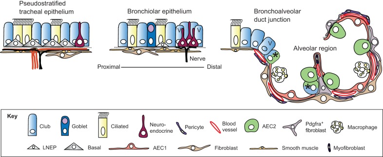
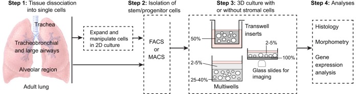
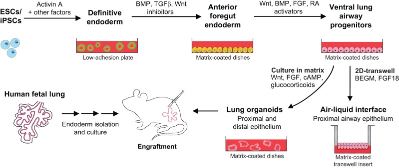
Lungs are composed of a system of highly branched tubes that bring air into the alveoli, where gas exchange takes place. The proximal and distal regions of the lung contain epithelial cells specialized for different functions: basal, secretory and ciliated cells in the conducting airways and type II and type I cells lining the alveoli. Basal, secretory and type II cells can be grown in three-dimensional culture, with or without supporting stromal cells, and under these conditions they give rise to self-organizing structures known as organoids. This Review summarizes the different methods for generating organoids from cells isolated from human and mouse lungs, and compares their final structure and cellular composition with that of the airways or alveoli of the adult lung. We also discuss the potential and limitations of organoids for addressing outstanding questions in lung biology and for developing new drugs for disorders such as cystic fibrosis and asthma.

Keywords: Lung organoids; Lung progenitors; Plasticity; Stem cells.


Conflict of interest statement

Competing interests

The authors declare no competing or financial interests.

Figures

Fig. 1.
Fig. 1.
Epithelial cell types of the mouse lung. Schematic of the major cell types in different regions of the mouse lung (Hogan et al., 2014). Goblet cells are much more abundant in human versus mouse airways. Basal cells expressing detectable levels of Trp63 and Krt5 are only present in trachea and main stem bronchi. Lineage-negative epithelial progenitors (LNEPs) have been proposed for the distal airways (Vaughan et al., 2015), which in the mouse are known as bronchioles as they lack associated cartilage. There is evidence that the Club cell population is heterogeneous, with a few cells in the bronchioalveolar duct junction (BADJ) and alveoli (marked with an asterisk) co-expressing Scbg1a1 and Sftpc (Kim et al., 2005; Rawlins et al., 2009). In addition, some Club cells in the vicinity of neuroendocrine bodies and the BADJ are resistant to killing by naphthalene. These ‘variant' Club cells (marked with V) can restore the population after damage (Giangreco et al., 2002). In the alveolar region, the two major epithelial cell types are type II (AEC2) and type I (AEC1) cells. The latter are closely apposed to capillary endothelial cells. Also present are a variety of stromal cells, including Pdgfra+ fibroblasts and lipofibroblasts (the latter located close to AEC2 cells), myofibroblasts and pericytes. Image modified from Rock and Hogan (2011).
Fig. 2.
Fig. 2.
Overview of the derivation of lung organoids. Cells isolated from different regions of the adult mouse and human lung have been used for 3D culture. If intact pieces of lung are used, rather than bronchial brushings for example, the tissue is dissociated using proteases (step 1). Primary cells are isolated using FACs or MACs (magnetic bead sorting) (step 2) and can be seeded directly into Matrigel (gray, percentage indicated). In the case of basal cells, the number of undifferentiated cells can be increased by culturing them in 2D before transferring to 3D. This enables genetic manipulation and the selection and cloning of specific mutants. Methods for expanding AEC2s in 2D have not yet been reported. In Step 3, single-cell suspensions are seeded into 3D culture in inserts or multiwells, with or without mesenchymal cells (Table 1). Methods include suspending the cells in 50% Matrigel (Rock et al., 2009) or in a low concentration of Matrigel and layering this over a higher concentration into which the cells sink (Butler et al., 2016; Danahay et al., 2015; Tata et al., 2013). For live imaging, cultures can be established in glass-bottomed wells coated with a thin layer of dense Matrigel. Cells sink through the upper layer and accumulate at the interface so that they remain in the same plane for imaging (Rock et al., 2011). For histological analysis, cultures are fixed in the Matrigel. For quantification of different cell types or passaging stem cells, the Matrigel can be removed using dispase and spheres dissociated with trypsin.
Fig. 3.
Fig. 3.
Basal cell-derived organoids. (A) Basal cell-derived organoids have been used for high- and medium-throughput screens (Danahay et al., 2015; Tadokoro et al., 2014). An example shows mouse tracheospheres after 7 days of culture, with and without 100 ng/ml noggin (a BMP inhibitor) and 20 ng/ml BMP4. The bottom right panel shows the results of scoring colony forming efficiency (CFE) in eight control wells versus eight wells with added noggin as one of the three values shown. The results were highly reproducible, giving a z factor of 1 (Zhang et al., 1999). It is important to establish such reproducibility before embarking on a large screen because conditions such as the position of a well in the tray, and changes in temperature and pH while changing the medium, can affect differentiation. (B) Schematic of a typical mouse tracheosphere after ∼14 days of culture, showing the relative position of basal versus luminal cells and markers of ciliated versus secretory cell types. (C) Section through clonal mouse tracheospheres cultured for 14 days and stained with antibody to Scgb3a2 (Club cells) and Foxj1 (ciliated cells). (D,D′) Sections of tracheospheres cultured without (D) or with (D′) 10 ng/ml IL13 and stained with DAPI and antibody to acetylated tubulin (cilia, red) and Splunc1 (Club cells, green). Note the dramatic increase in the number of secretory cells at the expense of ciliated cells in the presence of the cytokine. (E) Organoids (bronchospheres) derived from human basal cells cultured for 21 days without added factors (left), with the Rho kinase inhibitor Y-27632 (center), and with human lung fibroblasts (MRC5 line) (right). (F) Sections of human bronchospheres cultured for 21 days with MRC5 fibroblasts, stained with DAPI and markers for basal cells (KRT5, TRP63), luminal cells (KRT8, CLDN4), ciliated cells (FOXJ1) and secretory cells (MUC5AC). Scale bars: 100 µm in A insets, C,D′,F; 1 mm in A; 2 mm in E. Panel A was generated by Jason Rock; D,D′ by Tomomi Tadokoro.
Fig. 4.
Fig. 4.
AEC2-derived alveolosphere culture. (A) Sftpc-CreERT2/+; R26R-tdTomato/+; Pdgfra-GFP/+ mice were injected with tamoxifen, leading to lineage labeling of ∼80% of AEC2s (Tomato+). Arrows point to Pdgfra-GFP+ lipofibroblasts in close proximity to lineage-labeled AEC2s. (B) HTII280 is a surface marker coexpressed with SFTPC to mark AEC2s in human lung. (C) Lineage-labeled AEC2s and GFP+ fibroblasts from the mouse lung in A were isolated by FACS and placed into the alveolosphere culture system in a ratio of 1:10, respectively. The asterisk in the top panel and in the brightfield inset marks a large, lobular, non-lineage-labeled sphere that is likely to have derived from a non-AEC2 epithelial cell. Without the lineage label it would have been incorrectly assumed that this sphere derived from an AEC2. The bottom panel and higher magnification inset shows an example of several non-lineage-labeled alveolospheres (lacking a fluorescent signal) that are likely to be derived from AECs that had not undergone recombination of the reporter allele. (D) Section of an alveolosphere showing Sftpc+ AEC2s on the outside and AEC1s (Pdpn+) on the inside. (E) Section of an alveolosphere showing lineage-labeled (Tomato+) Ager-H2B:Venus+ AEC1s on the inside. Green cells that are not lineage labeled are Pdgfra-GFP+ stromal cells. (F) Schematic illustrating the main cellular components of an alveolosphere. Currently, the precise way in which the AEC2s and AEC1s are connected to each other is not known. (G) Lineage-labeled AEC2s can be isolated and passaged (at day 14) at least five times without significant loss of CFE. Scale bars: 25 μm in A; 50 μm in B,E; 500 μm in C.
Fig. 5.
Fig. 5.
Derivation of lung organoids from hPSCs. Directed differentiation protocols vary as to the components of the growth medium, the extracellular coating, and the stages at which the cells are placed in a 3D environment. The schematic is based on results from three groups (Huang et al., 2014, 2015; Dye et al., 2015, 2016; Wong et al., 2012) (see main text). Human pseudoglandular and canalicular stage (weeks 6-19 of gestation) fetal lungs can also provide an epithelial cell source. A combination of in vitro growth and subsequent in vivo engraftment currently provides the best conditions for maturation of lung epithelium. Culture of ventral lung progenitors in 2D air-liquid interface transwells generates only proximal conducting airway epithelium. RA, retinoic acid; BEGM, bronchial epithelial growth medium (Fulcher et al., 2005).

References

    1. Alder J. K., Barkauskas C. E., Limjunyawong N., Stanley S. E., Kembou F., Tuder R. M., Hogan B. L. M., Mitzner W. and Armanios M. (2015). Telomere dysfunction causes alveolar stem cell failure. Proc. Natl. Acad. Sci. USA 112, 5099-5104. 10.1073/pnas.1504780112 - DOI - PMC - PubMed
    1. Barkauskas C. E., Cronce M. J., Rackley C. R., Bowie E. J., Keene D. R., Stripp B. R., Randell S. H., Noble P. W. and Hogan B. L. M. (2013). Type 2 alveolar cells are stem cells in adult lung. J. Clin. Invest. 123, 3025-3036. 10.1172/JCI68782 - DOI - PMC - PubMed
    1. Butler C. R., Hynds R. E., Gowers K. H. C., Lee D. D. H., Brown J. M., Crowley C., Teixeira V. H., Smith C. M., Urbani L., Hamilton N. J. et al. (2016). Rapid expansion of human epithelial stem cells suitable for airway tissue engineering. Am. J. Respir. Crit. Care Med. 194, 156-168. 10.1164/rccm.201507-1414OC - DOI - PMC - PubMed
    1. Chen H., Matsumoto K., Brockway B. L., Rackley C. R., Liang J., Lee J.-H., Jiang D., Noble P. W., Randell S. H., Kim C. F. et al. (2012). Airway epithelial progenitors are region specific and show differential responses to bleomycin-induced lung injury. Stem Cells 30, 1948-1960. 10.1002/stem.1150 - DOI - PMC - PubMed
    1. Chu H. W., Rios C., Huang C., Wesolowska-Andersen A., Burchard E. G., O'Connor B. P., Fingerlin T. E., Nichols D., Reynolds S. D. and Seibold M. A. (2015). CRISPR-Cas9-mediated gene knockout in primary human airway epithelial cells reveals a proinflammatory role for MUC18. Gene Ther. 22, 822-829. 10.1038/gt.2015.53 - DOI - PMC - PubMed

Publication types