Review
. 2017 Jun;18(6):375-388.
doi: 10.1038/nrm.2017.11. Epub 2017 Mar 15.

Planar cell polarity in development and disease

Affiliations
Review

Planar cell polarity in development and disease

Mitchell T Butler et al. Nat Rev Mol Cell Biol. 2017 Jun.

Abstract

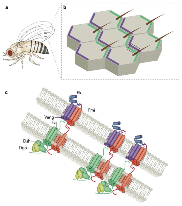
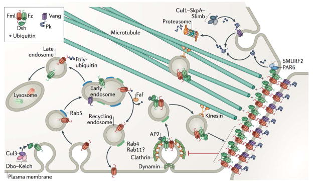
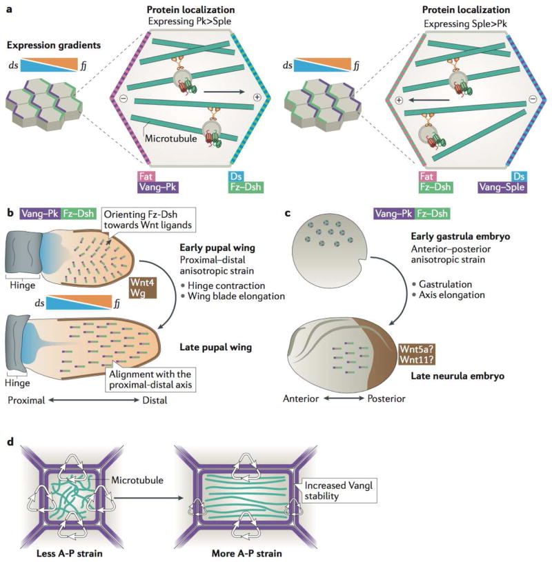
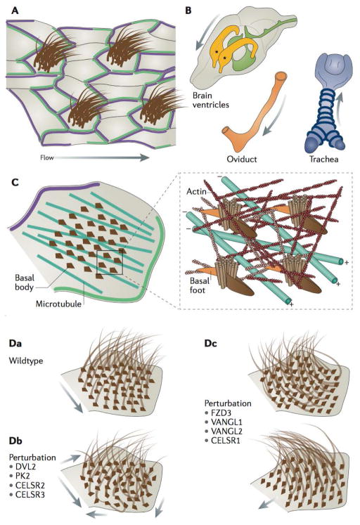
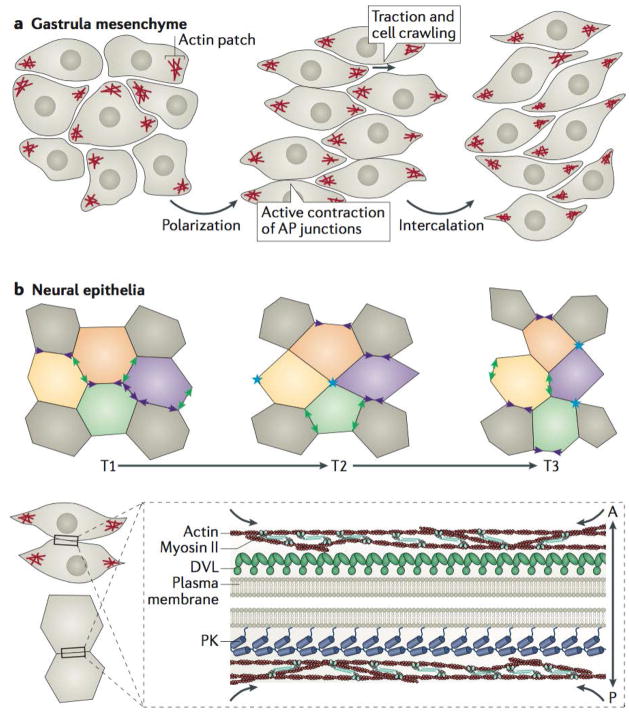
Planar cell polarity (PCP) is an essential feature of animal tissues, whereby distinct polarity is established within the plane of a cell sheet. Tissue-wide establishment of PCP is driven by multiple global cues, including gradients of gene expression, gradients of secreted WNT ligands and anisotropic tissue strain. These cues guide the dynamic, subcellular enrichment of PCP proteins, which can self-assemble into mutually exclusive complexes at opposite sides of a cell. Endocytosis, endosomal trafficking and degradation dynamics of PCP components further regulate planar tissue patterning. This polarization propagates throughout the whole tissue, providing a polarity axis that governs collective morphogenetic events such as the orientation of subcellular structures and cell rearrangements. Reflecting the necessity of polarized cellular behaviours for proper development and function of diverse organs, defects in PCP have been implicated in human pathologies, most notably in severe birth defects.


Figures

Figure 1
Figure 1. Asymmetric signalling complexes pattern planar polarity
a | Drosophila melanogaster has many planar-polarized external features, including actin-based, distally or posteriorly oriented hairs and bristles that cover the legs, wings, and notum, which serve as a robust readout of planar cell polarity signalling. The box drawn on the wing blade demarcates the region illustrated in (b). b | Cells of the D. melanogaster wing blade are asymmetrically patterned by proximal accumulations of Van Gogh (Vang) and Prickle (Pk) (both shown in purple) that are complementary to distal accumulations of Frizzled (Fz), Dishevelled (Dsh), and Diego (Dgo) (shown in in green). These patterns govern the distal positioning and orientation of a single actin-based trichome in each cell of the wing epithelium. The box drawn on the tissue demarcates the region illustrated in (c). c | Asymmetric PCP signalling components form junctional signalling complexes that are physically linked from cell to cell. Both Fz and Vang are transmembrane proteins that associate with physically linked Flamingo (Fmi) homodimers established between the opposing membranes at cell–cell junctions. Distal accumulations of Fmi and Fz are asymmetrically clustered through the activity of Dsh and Dgo, while proximal Fmi and Vang complexes are enriched by the activity of Pk. Note that the proteins as they are portrayed are not necessarily to scale and merely approximations as there is no high-resolution protein structural data available to support them.
Figure 2
Figure 2. Graphical summary of endocytosis, trafficking, and degradation events that facilitate the dynamic patterning of planar cell polarity
Planar cell polarity (PCP) signalling components are resistant to endocytic flux when stably linked across junctions (red inhibitory arrow),. The internalization of PCP components is partially dependent on dynamin and Clathrin-mediated endocytosis, and the interaction of membrane-associated Dishevelled (Dsh) with Clathrin-associated adaptor protein AP-2 is essential for PCP signalling. PCP protein internalization and trafficking are regulated through ubiquitylation. For example, the activity of a Cullin-3–Diablo–Kelch (Cul3–Dbo–Kelch) ubiquitin ligase complex promotes ubiquitylation and internalization – but not degradation – of Dsh, while fat facets (faf) deubiquitinase prevents the accumulation of Flamingo (Fmi) in Rab5-positive endosomes and promotes Fmi recycling to the membrane – a process that appears to be mediated by Rab4 and potentially Rab11 for PCP proteins. Although faf loss of function does not lead to a reduction in the overall levels of Fmi, inhibition of lysosomal maturation leads to the overaccumulation of Fmi as well as Frizzled (Fz) in endosomes, indicating that cellular levels of these components are, at least in part, regulated through lysosomal degradation. Kinesin-dependent trafficking of Fmi, Fz and Dsh towards microtubule plus ends appears to direct these complexes to sites of junctional enrichment. Prickle (Pk) appears to have a dual role in both clustering and stable Fmi–Van Gogh (Vang) complexes at junctions as well as in mediating the internalization of unstable Vang. Proteasomal degradation also plays a role in regulating PCP components. For example, Vang promotes the proteasomal degradation of farnesylated (membrane associated) Pk, likely through poly-ubiquitylation mediated by a Cullin-1 (Cul1)–SkpA–Supernumerary limbs (Slimb) E3 ubiquitin ligase complex. Similarly, vertebrate DVL2 (when phosphorylated in response to WNT5a stimulation) can associate with PAR6 and ubiquitin ligase SMURF2 resulting in poly-ubiquitylation of PK1, which leads to proteasomal degradation of PK1, thereby controlling PK1 localization and protein levels.
Figure 3
Figure 3. Multiple global inputs can collectively influence planar cell polarity orientation and stability across developing tissues
a | In Drosophila melanogaster core planar cell polarity (PCP) components organize according to Fat (Ft)–Dachsous (Ds) axis, established as a result of expression gradient of dachsous (ds) and the gradient of expression of kinase four-jointed (fj), which regulates the interactions between Ft and Ds and thereby contributes to the asymmetric distribution of these two proteins in the cell. Localization of PCP components with respect to Ft–Ds axis depends on the levels of expression of two Prickle isoforms: Prickle (Pk) and Spiny Legs (Sple). In cells expressing predominantly Pk, Vang–Pk localizes on the same side of the cell as Ft, but in cells expressing mostly Sple, Vang–Sple localizes together with Ds. This then regulates the directionality of microtubule-based transport in cells, as microtubule plus ends orient towards the Fz–Dsh-containing membranes. b | Top: The D. melanogaster wing blade exhibits a narrow source of Wnt ligands along the distal margin (brown) that may serve to orient PCP complex vectors (grey lines with Frizzled (Fz) and Dishevelled (Dsh) (green) localizing towards the wing margin opposite Vang Gogh (Vang) and Prickle (Pk) (purple) early in development (around 5-15 hours after puparium formation (hAPF)). Broad and shallow expression gradients of ds and fj oppose one another along the proximal–distal axis of the developing wing and may influence the directionality of early PCP pattern,, so that PCP is oriented in a radial fashion towards the edge of the wing. Bottom: Later in development (16–32 hAPF), during hinge contraction and rapid wing blade elongation, cell shape changes and cell rearrangement reorient microtubule polarity, leading to the coordinated alignment of asymmetric PCP patterns along the proximal–distal axis of the tissue (with Fz–Dsh oriented towards the distal edge of the tissue). Note the increased length of PCP vectors at the later stage that signifies an increase in the amount and stability of core PCP complexes at junctions c | Top: Prior to gastrulation in Xenopus laevis, no detectable PCP vector is observed (rings). Bottom: PCP axes (line vectors) are established in the embryonic ectoderm during gastrulation, as forces along the axis of elongation increase the cortical stability of Vang-like protein 2 (Vangl2) (purple)(see also panel d). A posterior source of Wnt ligands (such as Wnt5a and Wnt11;brown) is likely involved by directing the anterior localization of Vangl2. d | Anisotropic tissue strain orients microtubules along the axis of strain. Concomitantly, Vangl2 (purple) becomes more stable at cell–cell junctions along the axis of tissue strain, in a manner that has been shown to depend on microtubules.
Figure 4
Figure 4. Planar cell polarity patterns direct the cytoskeletal organization and ciliary polarity of multiciliated cells
a | Illustration of a multiciliated epithelium with asymmetric localization of planar cell polarity (PCP) complexes (green and purple), which direct polarized fluid flow across the surface of the tissue along the axis of PCP asymmetry (arrow). b | PCP patterning directs the fluid flow in various vertebrate tissues: the rostral flow of cerebrospinal fluid in the lateral ventricles (asterisks) of the brain, rostral mucociliary flow in the trachea, and medial flow in the oviduct of mice. c | PCP signalling is essential for the organization of the apical cytoskeletal elements responsible for ciliary orientation in multiciliated cells (MCCs),. An apical actin network (red) spaces basal bodies, whereas a subapical actin network (pink) links basal feet of one basal body to the neighboring basal body. These linkages to actin control rotational polarity, that ensures the uniform orientation of basal bodies, and thereby cilia in individual MCCs, whereas polarized apical microtubules orient groups of basal bodies along the axis of planar polarity,, with the microtubule plus-ends oriented towards Frizzled and Dishevelled (FZD–DVL) accumulations (green). d | Conserved rotational polarity observed in wild-type MCCs (a), can be disorganized by perturbation of DVL2, PK2, and CELSR2 and 3,,,, leading to largely disorganized cilia arrangements and disorganized ciliary beating and fluid flow (arrows) (b). Tissue-level polarity, the conservation of the uniform orientation of cilia across multiple MCCs, is disrupted upon perturbation of FZD3, Vang-like 1 (VANGL1) and VANGL2, and CELSR1,,; in this case different cells in the tissue can feature largely different orientation of their cilia. These defects can have considerable implications for the functions of MCCs in regulating fluid flow over tissues surfaces.
Figure 5
Figure 5. Planar cell polarity signalling directs polarized cell rearrangements during convergent extension
a | Mesenchymal cells during vertebrate gastrulation collectively elongate along the mediolateral axis, stabilize mediolateral actin-based protrusions, and mediolaterally intercalate to narrow and lengthen the tissue; all these processes require intact planar cell polarity (PCP) signalling,. b | Neural ectodermal cells mediolaterally intercalate by preferentially shrinking antero-posterior (AP) junctions (purple arrowheads), which first resolve (blue asterisks) and then elongate to form new medio-lateral (ML) junctions (green arrowheads). The stages of this intercalation behaviour are often referred to as “T” transitions, where T1 to T2 transitions involve the shrinking of the AP junction, whereas T2 to T3 transitions involve the lengthening of a new ML junction between two cells previously separated along the ML axis at T1. c | Core PCP components can be found asymmetrically enriched along AP junctions of cells undergoing convergent extension in vertebrates, with Prickle (PK) typically localizing to the anterior cell faces and Dishevelled (DVL) along the posterior cell faces. In the context of convergent extension, localization of PCP components at AP junctions coincides with the localization of filamentous actin enrichments and phosphorylated non-muscle Myosin II, which comprises the contractile actomyosin machinery that shrinks AP junctions,,,; there is evidence that PCP components are involved in regulating this contractility,.

References

    1. Goodrich LV, Strutt D. Principles of planar polarity in animal development. Development. 2011;138:1877–1892. - PMC - PubMed
    1. Adler PN. The frizzled/stan pathway and planar cell polarity in the Drosophila wing. Curr Top Dev Biol. 2012;101:1–31. - PMC - PubMed
    1. Peng Y, Axelrod JD. Asymmetric protein localization in planar cell polarity: Mechanisms, puzzles and challenges. Curr Top Dev Biol. 2012;101:33. - PMC - PubMed
    1. Lawrence PA, Casal J. The mechanisms of planar cell polarity, growth and the Hippo pathway: some known unknowns. Developmental Biology. 2013;377:1–8. - PMC - PubMed
    1. Yang Y, Mlodzik M. Wnt-Frizzled/Planar Cell Polarity Signalling: Cellular Orientation by Facing the Wind (Wnt) Annu Rev Cell Dev Biol. 2015;31:623–646. - PMC - PubMed

Publication types