

Proteomic profiling of fetal esophageal epithelium, esophageal cancer, and tumor-adjacent esophageal epithelium and immunohistochemical characterization of a representative differential protein, PRX6
- PMID: 28293090
- PMCID: PMC5330828
- DOI: 10.3748/wjg.v23.i8.1434
Proteomic profiling of fetal esophageal epithelium, esophageal cancer, and tumor-adjacent esophageal epithelium and immunohistochemical characterization of a representative differential protein, PRX6
Abstract
Aim: To understand the molecular mechanism of esophageal cancer development and provide molecular markers for screening high-risk populations and early diagnosis.
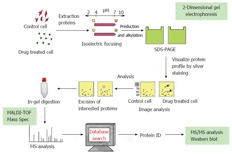
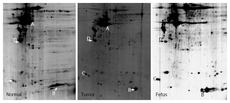
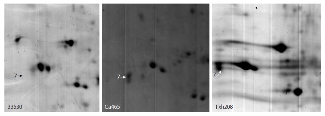
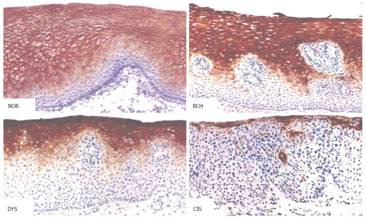
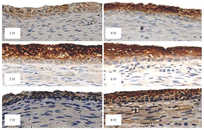
Methods: Two-dimensional electrophoresis combined with mass spectrometry were adopted to screen differentially expressed proteins in nine cases of fetal esophageal epithelium, eight cases of esophageal cancer, and eight cases of tumor-adjacent normal esophageal epithelium collected from fetuses of different gestational age, or esophageal cancer patients from a high-risk area of esophageal cancer in China. Immunohistochemistry (avidin-biotin-horseradish peroxidase complex method) was used to detect the expression of peroxiredoxin (PRX)6 in 91 cases of esophageal cancer, tumor-adjacent normal esophageal tissue, basal cell hyperplasia, dysplasia, and carcinoma in situ, as well as 65 cases of esophageal epithelium from fetuses at a gestational age of 3-9 mo.
Results: After peptide mass fingerprint analysis and search of protein databases, 21 differential proteins were identified; some of which represent a protein isoform. Varying degrees of expression of PRX6 protein, which was localized mainly in the cytoplasm, were detected in adult and fetal normal esophageal tissues, precancerous lesions, and esophageal cancer. With the progression of esophageal lesions, PRX6 protein expression showed a declining trend (P < 0.05). In fetal epithelium from fetuses at gestational age 3-6 mo, PRX6 protein expression showed a declining trend with age (P < 0.05). PRX6 protein expression was significantly higher in well-differentiated esophageal cancer tissues than in poorly differentiated esophageal cancer tissues (P < 0.05).
Conclusion: Development and progression of esophageal cancer result from interactions of genetic changes (accumulation or superposition). PRX6 protein is associated with fetal esophageal development and cancer differentiation.
Keywords: Esophageal squamous cell carcinoma; Fetal esophageal epithelium; Proteomics; Tumor-adjacent esophageal epithelium.
Conflict of interest statement
Conflict-of-interest statement: We have no financial relationships to disclose.
Figures
References
-
- Sheng KL, Zhu SY, Liu YS. Observation of human fetal eosphageal epihtelium. Acta Anat Sin. 1999;30:158–160.
-
- Johnson FP. The development of the mucous membrane of the esophagus, stomach and small intestine in the human embryo. Am J Anat. 1910;10:521–559.
-
- He QY, Chen J, Kung HF, Yuen AP, Chiu JF. Identification of tumor-associated proteins in oral tongue squamous cell carcinoma by proteomics. Proteomics. 2004;4:271–278. - PubMed
-
- Srisomsap C, Sawangareetrakul P, Subhasitanont P, Panichakul T, Keeratichamroen S, Lirdprapamongkol K, Chokchaichamnankit D, Sirisinha S, Svasti J. Proteomic analysis of cholangiocarcinoma cell line. Proteomics. 2004;4:1135–1144. - PubMed
-
- Wang LD, Shi ST, Zhou Q, Goldstein S, Hong JY, Shao P, Qiu SL, Yang CS. Changes in p53 and cyclin D1 protein levels and cell proliferation in different stages of human esophageal and gastric-cardia carcinogenesis. Int J Cancer. 1994;59:514–519. - PubMed
MeSH terms
Substances
LinkOut - more resources
Full Text Sources
Other Literature Sources
Medical
Miscellaneous
