. 2017 Feb 28:8:303.
doi: 10.3389/fmicb.2017.00303. eCollection 2017.

MLKL Mediated Necroptosis Accelerates JEV-Induced Neuroinflammation in Mice

Affiliations

MLKL Mediated Necroptosis Accelerates JEV-Induced Neuroinflammation in Mice

Peiyu Bian et al. Front Microbiol. .

Abstract

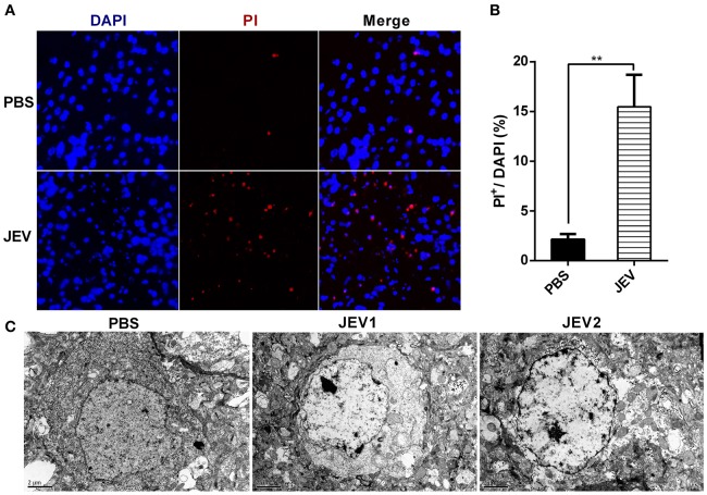
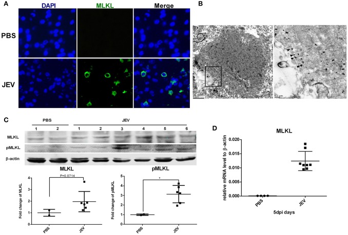
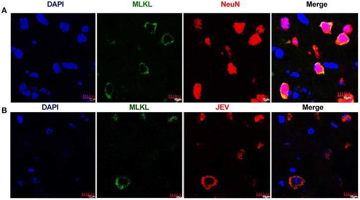
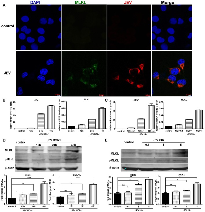
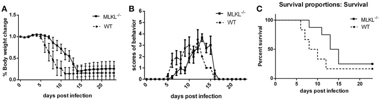
Japanese encephalitis virus (JEV) is the most prevalent cause of viral encephalitis in Asia and the western Pacific. Neuronal death caused by JEV infection and inflammation induced cytotoxicity leads to progression and deterioration of Japanese encephalitis (JE). Mixed-lineage kinase domain-like protein (MLKL) mediated necroptosis is a newly discovered pathway of programmed cell death and participates in many inflammatory diseases. In this study, we demonstrated for the first time that necroptosis was involved in the neuronal loss during JE via immune-electron microscopy and immunochemistry. The expression of MLKL in neurons was upregulated in presence of JEV infection in vitro and in vivo. Deletion of MLKL alleviated the progression of JE and decreased the level of inflammatory cytokines in mice model. Taken together, this study provides evidence for the participation of necroptosis in the pathogenesis of JEV infection.

Keywords: Japanese encephalitis virus; MLKL; inflammation; necroptosis; neuronal death.


Figures

Figure 1
Figure 1
Nerve cells undergo necrosis during JE in mice model. C57BL/6 mice were infected i.p. with PBS or JEV 5 × 10⁷ PFU in 200 μl PBS/20 g per mouse. From 5th dpi, JEV infected mice developed body weight loss and early clinical signs such as piloerection, and physical limitation. Brains from each mouse were harvested for further experiment at 5 dpi. (A) The representative images of PI staining of brain sections from PBS or JEV administrated mice (200x). (B) The intensity of PI positively stained cells of each group were analyzed with Image J. (Data represents mean ± SEM. PBS = 2, JEV = 3; 3 sections per mouse, 5 fields per section, **P < 0.01). (C) Micrographs of normal and JEV infected mouse brain cells. In PBS group, the morphology of brain cells. In JEV infected mice, most of brain cells showed classical necrotic morphology with clumps of chromatin, swollen mitochondria and plasma membrane disintegration (PBS = 1, JEV = 2).
Figure 2
Figure 2
MLKL mediated necroptosis is involved in JE. Mice were infected i.p. with PBS or JEV 5 × 10⁷ PFU in 200 μl PBS/20 g. At 5 dpi, brains were harvested for immunochemistry, immunoelectromicroscopy, westeron blot and qRT-PCR. (A) The immunochemistry of MLKL in the brain sections of PBS or JEV administered mice (x400). There was obvious staining of MLKL around the plasma membrane in the brain sections of JEV infected mice while it was invisible in control mice. (B) Immuno-electron microscopic study of MLKL. The right panels show magnified regions of the boxed areas. Arrows showed the membrane localization of MLKL. (C) Western-blotting (upper) and quantitation (down) of protein MLKL and pMLKL in PBS or JEV administrated group. There was increased expression of protein MLKL and pMLKL in JEV infected group (PBS = 2, JEV = 6, *P < 0.05). (D) The relative level of mRNA MLKL in PBS or JEV treated mice brain via qRT-PCR at 5 dpi. The data represents the relative mRNA level normalized with β-actin (PBS = 4, JEV = 8). The mRNA MLKL was significantly increased in JEV infected mouse brains compared with PBS group.
Figure 3
Figure 3
Necroptosis of neurons after JEV infection. The double-staining of MLKL and NeuN as well as the double-staining of JEV and MLKL was conducted in brain sections from JEV infected mice at 5 dpi. (A) Double-immunostaining of MLKL (green) and NeuN (red) in JEV infected mice. The results were acquired by confocal laser scanning microscope. The increased expression of MLKL was mainly occurred in neurons. (B) Double-immunostaining of MLKL (green) and JEV (red) in JEV infected mice. The expression of MLKL was closely correlated with JEV infection.
Figure 4
Figure 4
The expression of MLKL is upregulated in neurons infected with JEV. The expression of MLKL in Neuro2a cells was detected through immunofluorescence after JEV infection. Meanwhile, the mRNA level of MLKL and protein level of MLKL and pMLKL in Neuro2a cells were tested through qRT-PCR and western blotting after JEV infection at different MOI and infection time (The data represents the mean ± SEM for 3 independent experiments, *P < 0.05, **P < 0.01, ***P < 0.001 and ****P < 0.0001). (A) Double-immunostaining of MLKL (green) and JEV (red) in Neuro2a cells treated with DMEM (top) and infected with JEV MOI = 1, 24 h (bottom). (B) Neuro2a cells were infected with JEV at MOI = 1. And 12, 24, 48 h later, cells were collected for RNA extraction and qRT-PCR. The viral copies and the level of mRNA MLKL increased as the extension of infection time. (C) Neuro2a cells were infected with JEV at MOI = 0.1, 1, 5 respectively. And 24 h later, cells were collected for RNA extraction and qRT-PCR. The viral copies and the level of mRNA MLKL increased as the increase of infection dose. (D) Neuro2a cells were infected with JEV at MOI = 1. And 12, 24, 48 h later, cells were collected for total protein extraction and western-bloting. With the extension of infection time, the expression of protein MLKL and pMLKL increased. (E) Neuro2a cells were infected with JEV at MOI = 0.1, 1, 5 respectively. And 24 h later, cells were collected for total protein extraction and western-bloting. The level of protein MLKL and pMLKL increased as the increase of infection dose.
Figure 5
Figure 5
MLKL deleted mice show alleviated progression of JE. Wild and MLKL−/− mice were administered with JEV 5 × 10⁷ PFU/20 g in 200 μl PBS intraperitoneally. The body weight, behavior scores and survival rate were recorded daily from 0 dpi to 23 dpi when the survivals were completely stable (WT = 12, MLKL−/− = 16). (A) The weight of each mouse was measured and recorded at 16: 30–17: 00 (the data represents the mean ± SD). Compared with wild mice, MLKL−/− group showed alleviated weight loss. (B) The behavior score was recorded twice every day according to the scoring criteria (0 = no piloerection, no restriction of movement, no body stiffening, no hind limb paralysis. 1 = piloerection, no restriction of movement, no body stiffening, no hind limb paralysis. 2 = piloerection, restriction of movement, no body stiffening, no hind limb paralysis. 3 = piloerection, restriction of movement, body stiffening, no hind limb paralysis. 4 = piloerection, restriction of movement, body stiffening, hind limb paralysis. 5 = piloerection, restriction of movement, body stiffening, hind limb paralysis, sometimes tremor even death. And the dead mice were excluded from the cohort and the mean scores of all mice in each group were calculated. The data represent the mean ± SD). MLKL−/− mice showed slowed onset and progression of JE compared with wild group. (C) The death and survival of mice in each group was recorded and the data was analyzed and shown as Kaplan–Meier survival curves. Even though there was no significant difference in the final survival rate, MLKL−/− group survived for longer time than wild group.
Figure 6
Figure 6
MLKL deleted mice show decreased inflammatory cytokines during JEV infection. The serum and brain from each mice in MLKL−/− and wild group were collected at 5 dpi, and the main inflammatory cytokines in serum and brain were tested. (A) The level of CCL-2, IL-1β, IFN-γ, TNF-α in the serum of WT or MLKL−/− mice was detected by ELISA assay kit (WT-PBS = 2, MLKL−/−-PBS = 2, WT-JEV = 9, MLKL−/−-PBS = 9). (B) The expression of CCL-2, IL-1β, IFN-γ, TNF-α in the brain of WT or MLKL−/− mice was detected by qRT-PCR (WT-PBS = 2, MLKL−/−-PBS = 2, WT-JEV = 9, MLKL−/−-PBS = 9, *P < 0.05, ***P < 0.001).

References

    1. Biswas S. M., Kar S., Singh R., Chakraborty D., Vipat V., Raut C. G., et al. . (2010). Immunomodulatory cytokines determine the outcome of Japanese encephalitis virus infection in mice. J. Med. Virol. 82, 304–310. 10.1002/jmv.21688 - DOI - PubMed
    1. Cai Z., Jitkaew S., Zhao J., Chiang H. C., Choksi S., Liu J., et al. . (2014). Plasma membrane translocation of trimerized MLKL protein is required for TNF-induced necroptosis. Nat. Cell Biol. 16, 55–65. 10.1038/ncb2883 - DOI - PMC - PubMed
    1. Chen C. J., Ou Y. C., Chang C. Y., Pan H. C., Liao S. L., Chen S. Y., et al. . (2012). Glutamate released by Japanese encephalitis virus-infected microglia involves TNF-α signaling and contributes to neuronal death. Glia 60, 487–501. 10.1002/glia.22282 - DOI - PubMed
    1. Chen C. J., Ou Y. C., Lin S. Y., Raung S. L., Liao S. L., Lai C. Y., et al. . (2010). Glial activation involvement in neuronal death by Japanese encephalitis virus infection. J. Gen. Virol. 91, 1028–1037. 10.1099/vir.0.013565-0 - DOI - PubMed
    1. Dhib-Jalbut S., Kalvakolanu D. V. (2015). Microglia and necroptosis: the culprits of neuronal cell death in multiple sclerosis. Cytokine 76, 583–584. 10.1016/j.cyto.2015.06.004 - DOI - PubMed

LinkOut - more resources