

[The role of dermoscopy in the diagnosis of basosquamous carcinoma]
- PMID: 28293368
- PMCID: PMC5337263
- DOI: 10.11604/pamj.2016.25.252.11345
[The role of dermoscopy in the diagnosis of basosquamous carcinoma]
Abstract
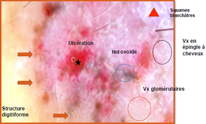
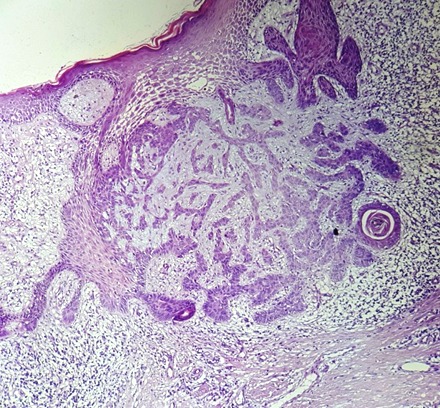
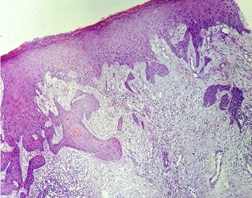
Basosquamous carcinoma (BSC) is a rare skin cancer which has areas of basal cell carcinoma (BCC) and squamous cell carcinoma (SCC) and a transition zone between them. However, dermoscopic features of BSC are not well described in the literature, except for two studies. The aim of this case study was to better identify and clarify the role of dermoscopy in the diagnosis of BSC, although histological confirmation is necessary.
Le carcinome basosquameux (CBS) est un cancer cutané rare qui présente des zones de carcinome basocellulaire (CBC) et de carcinome épidermoïde (SCC) et une zone de transition entre elles. Cependant, les caractéristiques dermoscopiques du BSC ne sont pas bien décrites dans la littérature, sauf deux études. Le but du présent cas était de mieux identifier et clarifier la contribution de la dermoscopie dans le diagnostic du BSC, malgré que la confirmation reste toujours histologique.
Keywords: Basosquamous carcinoma; basal cell carcinoma; dermoscopy; squamous cell carcinoma.
Conflict of interest statement
Les auteurs ne déclarent aucun conflits d’intérêts.
Figures

References
-
- Giacomel J, Lallas A, Argenziano G, Reggiani C, Piana S, Apalla Z, Ferrara G, Moscarella E, Longo C, Zalaudek I. Dermoscopy of basosquamous carcinoma. Br J Dermatol. 2013 Aug;169(2):358–64. - PubMed
-
- Akay BN, Saral S, Heper AO, Erdem C, Rosendahl C. Basosquamous carcinoma: dermoscopic clues to diagnosis. J Dermatol. 2016 Aug 29;169(2):358–64. - PubMed
-
- Boyd AS, Rapini RP. Cutaneous collision tumors: an analysis of 69 cases and review of the literature. Am J Dermatopathol. 1994;16(3):253–7. - PubMed
-
- Zaballos P, Llambrich A, Puig S, Malvehy J. Dermoscopy is useful for the recognition of benign-malignant compound tumors. Br J Dermatol. 2005;15(3):653–6. - PubMed
-
- Moscarella E, Rabinovitz H, Oliviero MC, Brown L, Longo C, Zalaudek I, et al. The role of reflectance confocal microscopy as an aid in the diagnosis of collision tumors. Dermatology. 2013;227(2):109–17. - PubMed
Publication types
MeSH terms
LinkOut - more resources
Full Text Sources
Other Literature Sources
Medical
Research Materials
