Effect of Axonal Trauma on Nerve Regeneration in Side-to-side Neurorrhaphy: An Experimental Study
- PMID: 28293524
- PMCID: PMC5222669
- DOI: 10.1097/GOX.0000000000001180
Effect of Axonal Trauma on Nerve Regeneration in Side-to-side Neurorrhaphy: An Experimental Study
Abstract
Background: Side-to-side (STS) neurorrhaphy can be performed distally to ensure timely end-organ innervation. It leaves the distal end of the injured nerve intact for further reconstruction. Despite encouraging clinical results, only few experimental studies have been published to enhance the regeneration results of the procedure. We examined the influence of different size epineural windows and degree of axonal injury of STS repair on nerve regeneration and donor nerve morbidity.
Methods: Three clinically relevant repair techniques of the transected common peroneal nerve (CPN) were compared. Group A: 10-mm long epineural STS windows; group B: 2-mm long windows and partial axotomy to the donor tibial nerve; and group C: 2-mm long windows with axotomies to both nerves. Regeneration was followed by the walk track analysis, nerve morphometry, histology, and wet muscle mass calculations.
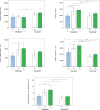
Results: The results of the walk track analysis were significantly better in groups B and C compared with group A. The nerve fiber count, total fiber area, fiber density, and percentage of the fiber area values of CPN of the group C were significantly higher when compared with group A. The wet mass ratio of the CPN-innervated anterior tibial muscle was significantly higher in group C compared with group A. The wet mass ratio of the tibial nerve-innervated gastrocnemial muscle was higher in group A compared with the other groups.
Conclusions: All three variations of the STS repair technique showed nerve regeneration. Deliberate donor nerve axotomy enhanced nerve regeneration. A larger epineural window did not compensate the effect of axonal trauma on nerve regeneration.
Figures


Similar articles
-
Protective distal side-to-side neurorrhaphy in proximal nerve injury-an experimental study with rats.Acta Neurochir (Wien). 2019 Apr;161(4):645-656. doi: 10.1007/s00701-019-03835-2. Epub 2019 Feb 12. Acta Neurochir (Wien). 2019. PMID: 30746570 Free PMC article.
-
End-to-Side vs. Free Graft Nerve Reconstruction-Experimental Study on Rats.Int J Mol Sci. 2023 Jun 21;24(13):10428. doi: 10.3390/ijms241310428. Int J Mol Sci. 2023. PMID: 37445608 Free PMC article.
-
Efficacy of the end-to-side neurorrhaphies with epineural window and partial donor neurectomy in peripheral nerve repair: an experimental study in rats.J Reconstr Microsurg. 2015 Jan;31(1):31-8. doi: 10.1055/s-0034-1382263. Epub 2014 Aug 1. J Reconstr Microsurg. 2015. PMID: 25083764
-
End-to-side nerve neurorrhaphy: critical appraisal of experimental and clinical data.Acta Neurochir Suppl. 2007;100:77-84. doi: 10.1007/978-3-211-72958-8_17. Acta Neurochir Suppl. 2007. PMID: 17985551 Review.
-
Chapter 13: Experimental results in end-to-side neurorrhaphy.Int Rev Neurobiol. 2009;87:269-79. doi: 10.1016/S0074-7742(09)87013-X. Int Rev Neurobiol. 2009. PMID: 19682642 Review.
Cited by
-
Molecular Basis of Surgical Coaptation Techniques in Peripheral Nerve Injuries.J Clin Med. 2023 Feb 16;12(4):1555. doi: 10.3390/jcm12041555. J Clin Med. 2023. PMID: 36836090 Free PMC article. Review.
References
-
- Yüksel F, Karacaoğlu E, Güler MM. Nerve regeneration through side-to-side neurorrhaphy sites in a rat model: a new concept in peripheral nerve surgery. Plast Reconstr Surg. 1999;104:2092–2099. - PubMed
-
- Yüksel F, Peker F, Celiköz B. Two applications of end-to-side nerve neurorrhaphy in severe upper-extremity nerve injuries. Microsurgery. 2004;24:363–368. - PubMed
-
- Cage TA, Simon NG, Bourque S, et al. Dual reinnervation of biceps muscle after side-to-side anastomosis of an intact median nerve and a damaged musculocutaneous nerve. J Neurosurg. 2013;119:929–933. - PubMed
LinkOut - more resources
Full Text Sources
Other Literature Sources
