. 2017 Feb 28:7:56.
doi: 10.3389/fcimb.2017.00056. eCollection 2017.

Induction of Atypical Autophagy by Porcine Hemagglutinating Encephalomyelitis Virus Contributes to Viral Replication

Affiliations

Induction of Atypical Autophagy by Porcine Hemagglutinating Encephalomyelitis Virus Contributes to Viral Replication

Ning Ding et al. Front Cell Infect Microbiol. .

Abstract

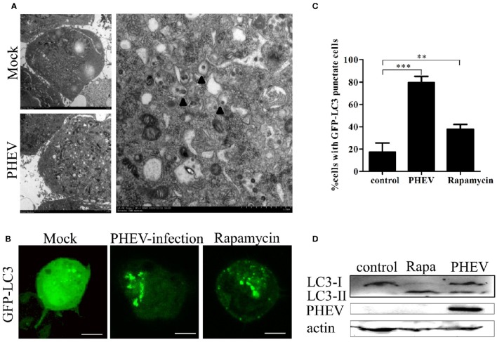
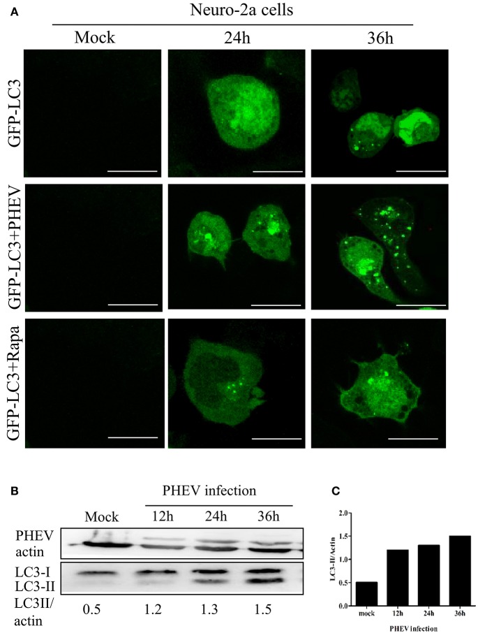
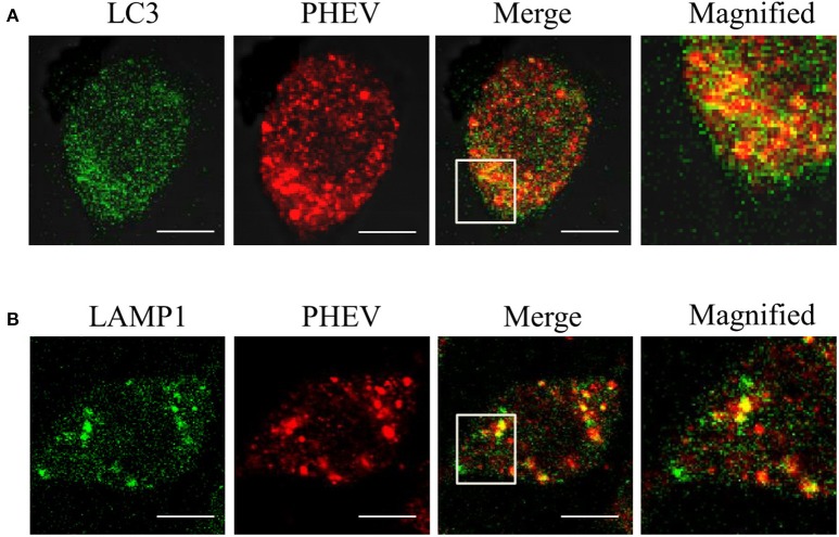
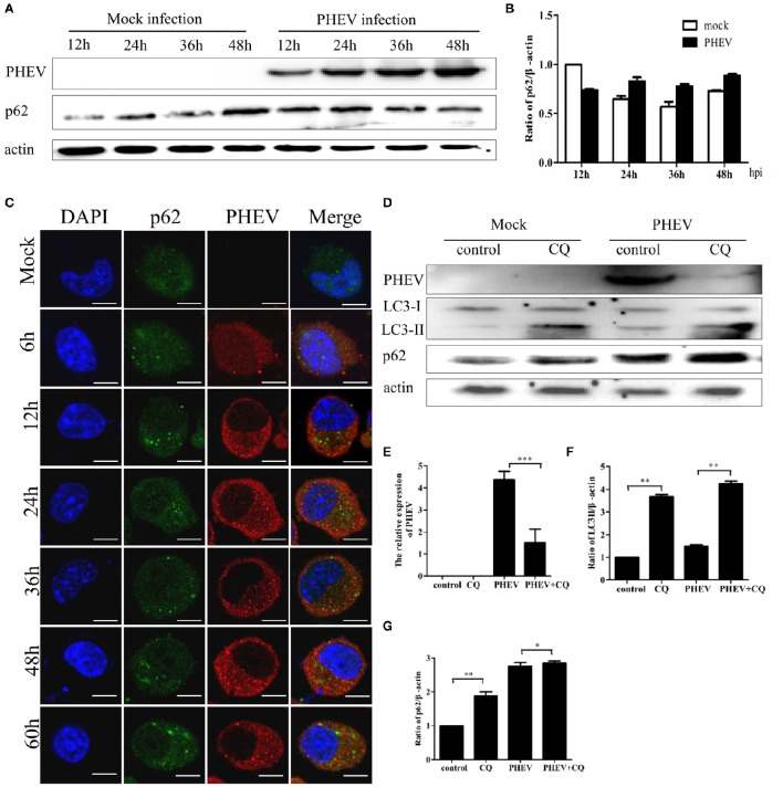
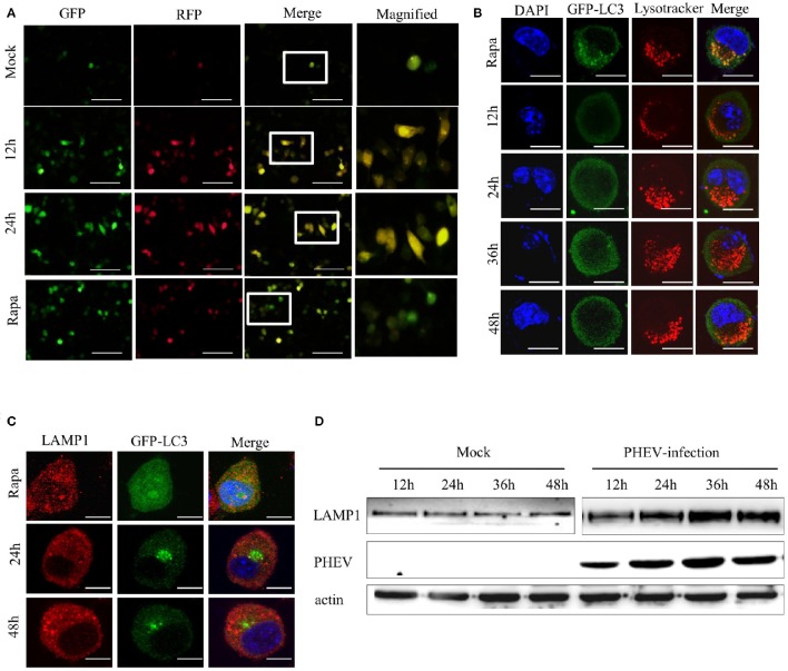
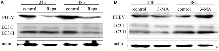
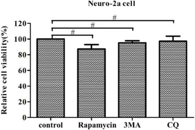
Autophagy is a basic biological metabolic process involving in intracellular membrane transport pathways that recycle cellular components and eliminate intracellular microorganisms within the lysosome. Autophagy also plays an important part in virus infection and propagation. However, some pathogens, including viruses, have evolved unique trick to escape or exploit autophagy. This study explores the mechanism of autophagy induction by porcine hemagglutinating encephalomyelitis virus (PHEV) in Neuro-2a cells, and examines the role of autophagy in PHEV replication. PHEV triggered autophagy in Neuro-2a cells is dependent on the presence of bulk double- or single-membrane vacuoles, the accumulation of GFP-LC3 fluorescent dots, and the LC3 lipidation. In addition, PHEV induced an incomplete autophagic effect because the degradation level of p62 did not change in PHEV-infected cells. Further validation was captured using LysoTracker and lysosome-associated membrane protein by indirect immunofluorescence labeling in PHEV-infected cells. We also investigated the change in viral replication by pharmacological experiments with the autophagy inducer rapamycin or the autophagy inhibitor 3-MA, and the lysosomal inhibitor chloroquine (CQ). Suppression of autophagy by 3-MA increased viral replication, compared with the mock treatment, while promoting of autophagy by rapamycin reduced PHEV replication. CQ treatment enhanced the LC3 lipidation in PHEV-infected Neuro-2a cells but lowered PHEV replication. These results show that PHEV infection induces atypical autophagy and causes the appearance of autophagosomes but blocks the fusion with lysosomes, which is necessary for the replication of PHEV in nerve cells.

Keywords: LC3; atypical autophagy; autophagic flux; neuro-2a cells; neurotropic virus; porcine hemagglutinating encephalomyelitis virus; virus replication.


Figures

Figure 1
Figure 1
Autophagosomes accumulate in PHEV-infected Neuro-2a cells. (A) TEM observations. Neuro-2a cells were mock infected or infected with PHEV for 24 h and studied by transmission electron microscopy. Black triangles indicate the structures with autophagosomes characteristics. (B) Confocal microscopy. Neuro-2a cells were transfected with GFP-LC3 followed by treatment at 24 h post transfection with mock treatment as a negative control, and rapamycin treatment as a positive control. The localization of GFP-LC3 positive autophagosome accumulation (green) and the S-tagged PHEV products (red) was visualized using a confocal microscope. (C) The quantification of cells showing GFP-LC3 puncta in PHEV-infected cells. In three random fields, the average number of puncta in each cell was taken from at least 80 cells in each treatment. Representative results with graphs are shown in Figure 1B. (D) Western blotting. The turnovers of LC3-I to LC3-II were detected for mock-treated Neuro-2a cells, rapamycin-treated Neuro-2a cells, and PHEV-infected Neuro-2a cells. Cells were collected at appropriate time points and detected with anti-LC3B antibody, and β-actin was used as a protein loading control. **P < 0.01; ***P < 0.001. Scale bars: 10 μm.
Figure 2
Figure 2
PHEV-induced autophagy was time-dependent. (A) Immunofluorescence microscopy was used to detect the GFP-LC3-autophagosomes in PHEV infected cells at different post-infection time points before transfection with GFP-LC3 into Neuro-2a cells. The cells were fixed at 0, 24, and 36 h post-infection, respectively, and then analyzed for GFP-LC3 positive autophagosome accumulation using a confocal microscope as described in Figure 2A. (B,C) Neuro-2a cells were infected with PHEV and mock-infected cells served as control. At 0, 12, 24, and 36 h post-infection, the cells were then lysed for western blotting analysis using an anti-LC3B and anti-β-actin antibodies. Scale bars: 10 μm.
Figure 3
Figure 3
The membranes of autophagosome-like vesicles have colocalization with virions. (A,B) PHEV-infected Neuro2a cells were stained with LC3 (green) and PHEV (red) antibodies. The color-merged images are shown in the third panel. The colocalization between PHEV and LC3 and LAMP1 was magnified in the merged images. Scale bars: 10 μm.
Figure 4
Figure 4
PHEV infection suppresses autophagic flux. (A) Western blot analysis of p62 changes in Neuro-2a cells with PHEV infection for 12, 24, 36, and 48 h, as well as the mock infection for the relevant times. (B) The p62 to β-actin ratio normalized to the mock infection set at 1.0 of (A) (n = 3; P < 0.05). (C) Representative images of Neuro-2a cells with PHEV infection from 6 to 60 h labeled with antibodies to anti-p62 and anti-PHEV. (D) Effect of chloroquine (CQ) treatment on LC3-II and p62 of Neuro-2a cells for 24 h, as tested by Western blotting. (E–G) show the ratios of PHEV to β-actin, p62 to β-actin or LC3-II to β-actin from three independent experiments of (D). *P < 0.05; **P < 0.01; ***P < 0.001. Scale bars: 10 μm.
Figure 5
Figure 5
PHEV activates autophagosome formation but prevents its fusion with lysosomes. (A) Neuro-2a cells were transfected with mRFP-GFP-LC3. As the positive control for induction of autophagy, Neuro-2a cells were transfected with mRFP-GFP-LC3 and then treated with complete medium supplemented with 200 μM rapamycin for 3 h. At 0, 12, and 24 h post-transfection, the cells were fixed and assessed with GFP and mRFP fluorescence. Scale bars: 50 μm. (B) Neuro-2a cells were used to analyze the colocalization of LysoTracker-stained acidified vesicles and GFP-LC3-positive autophagosomes in the mock-infected and PHEV-infected cells at 12, 24, 36, and 48 h. Representative images are shown in Figure 5B. (C) Furthermore, the fusion of autophagosomes with lysosomes was analyzed as colocalization of the autophagosome marker GFP-LC3 with the lysosome marker LAMP1. Nuclear DNA was stained with DAPI. One of the three experiments conducted is shown. (D) Western blotting analysis of LAMP1 changes in PHEV-infected cells for 12, 24, 36, and 48 h were shown in Figure 5D, compared with the relevant times in mock cells. Scale bars: 10 μm.
Figure 6
Figure 6
The effect of -induced or -inhibited autophagy on PHEV replication. (A,B) Neuro-2a cells were infected with PHEV in the presence or absence of rapamycin (100 nM) or 3-MA (2 mM). At 24 h and 48 hpi, the cells were harvested and analyzed by immunoblotting using anti-LC3, anti-PHEV, and anti-β-actin antibodies.
Figure 7
Figure 7
Regulation of autophagy does not affect cell viability. The pharmacological alteration of autophagy does not affect cell viability. Neuro-2a cells were determined by WST-8 cell proliferation assay after being treated with rapamycin, 3-MA, or chloroquine (CQ) for 24 h. Data on the percentage of cell viability are shown as the mean ± SD for three independent experiments. #P > 0.05.

Similar articles

Cited by

References

    1. Andries K., Pensaert M. (1981). Vomiting and wasting disease, a coronavirus infection of pigs. Adv. Exp. Med. Biol. 142, 399–408. 10.1007/978-1-4757-0456-3_33 - DOI - PubMed
    1. Andries K., Pensaert M. B. (1980). Immunofluorescence studies on the pathogenesis of hemagglutinating encephalomyelitis virus infection in pigs after oronasal inoculation. Am. J. Vet. Res. 41, 1372–1378. - PubMed
    1. Beachboard D. C., Anderson-Daniels J. M., Denison M. R. (2015). Mutations across murine hepatitis virus nsp4 alter virus fitness and membrane modifications. J. Virol. 89, 2080–2089. 10.1128/JVI.02776-14 - DOI - PMC - PubMed
    1. Bjørkøy G., Lamark T., Brech A., Outzen H., Perander M., Overvatn A., et al. . (2005). p62/SQSTM1 forms protein aggregates degraded by autophagy and has a protective effect on huntingtin-induced cell death. J. Cell Biol. 171, 603–614. 10.1083/jcb.200507002 - DOI - PMC - PubMed
    1. Blanchard E., Roingeard P. (2015). Virus-induced double-membrane vesicles. Cell. Microbiol. 17, 45–50. 10.1111/cmi.12372 - DOI - PMC - PubMed

Publication types

MeSH terms

LinkOut - more resources