. 2017:2017:1857580.
doi: 10.1155/2017/1857580. Epub 2017 Feb 15.

Integrating miRNA and mRNA Expression Profiling Uncovers miRNAs Underlying Fat Deposition in Sheep

Affiliations

Integrating miRNA and mRNA Expression Profiling Uncovers miRNAs Underlying Fat Deposition in Sheep

Guangxian Zhou et al. Biomed Res Int. 2017.

Abstract

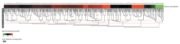
MicroRNAs (miRNAs) are endogenous, noncoding RNAs that regulate various biological processes including adipogenesis and fat metabolism. Here, we adopted a deep sequencing approach to determine the identity and abundance of miRNAs involved in fat deposition in adipose tissues from fat-tailed (Kazakhstan sheep, KS) and thin-tailed (Tibetan sheep, TS) sheep breeds. By comparing HiSeq data of these two breeds, 539 miRNAs were shared in both breeds, whereas 179 and 97 miRNAs were uniquely expressed in KS and TS, respectively. We also identified 35 miRNAs that are considered to be putative novel miRNAs. The integration of miRNA-mRNA analysis revealed that miRNA-associated targets were mainly involved in the gene ontology (GO) biological processes concerning cellular process and metabolic process, and miRNAs play critical roles in fat deposition through their ability to regulate fundamental pathways. These pathways included the MAPK signaling pathway, FoxO and Wnt signaling pathway, and focal adhesion. Taken together, our results define miRNA expression signatures that may contribute to fat deposition and lipid metabolism in sheep.


Conflict of interest statement

The authors declare that they have no competing interests.

Figures

Figure 1
Figure 1
(a) Length distribution of the clean reads based on total abundance and distinct sequences. (b) Differential expression of miRNAs between the adipose tissues from KS and TS breed. Each point represents a single miRNA. The X and Y axes show the expression level of miRNAs in these two samples, respectively. Red points represent miRNAs with a ratio > 2, blue points represent miRNAs with 1/2 < ratio ≦ 2, and green points represent miRNAs with ratio ≦ 1/2; ratio = normalized expression in treatment/normalized expression in control.
Figure 2
Figure 2
Heat map of miRNAs differentially expressed in sheep adipose tissues. Red indicates that the miRNA has a higher expression level in the treatment samples; green indicates that the miRNA has a higher expression in the control samples and gray indicates that the miRNA has no expression in at least one sample. Each row in the figure represents one miRNA, and each column shows one sample pair. Each cell shows the differential expression of a miRNA in one sample pair.
Figure 3
Figure 3
qPCR validation of miRNAs identified in sheep adipose tissues. Red represents TS and blue represents KS (0.01 < p < 0.05, ∗∗p < 0.01).
Figure 4
Figure 4
Differentially expressed gene between TS and KS DEGs. Red spots represented upregulated genes, and green spots indicated downregulated genes. Back spots represented genes that did not show obvious changes between the TS and KS.
Figure 5
Figure 5
GO classification of DEG. The x-axis indicated the subcategories; the right y-axis indicated the number of DEG. The height of columns represented the percentage (number) of upregulated expression (red) and downregulated expression (green) genes.
Figure 6
Figure 6
Top 20 of pathway enrichment. The circle size represented gene number. Q-value was shown by color gradient.
Figure 7
Figure 7
miR-125a-5p downregulates the expression of ESRRα by targeting its 3′ UTR site. (a) The pairing schematic of ESRRα 3′ UTR and miR-125a-5p. The nucleotide in blue represents “seed sequence” of miR-125a-5p, and the mutation nucleotides are presented in red. (b) The insertion site of ESRRα 3′ UTR or its mutation luciferase reporter vector map. (c) The two types were cotransfected with miR-125a-5p mimics (or negative control) into 293T cells. Luciferase assay was performed 48 h after transfection. Results are presented as relative luciferase activity (mean ± SD; n = 4; ANOVA; ∗∗P < 0.01).

Similar articles

Cited by

References

    1. Davidson A. Oxford Companion to Food. Oxford, UK: Oxford University Press; 1999.
    1. Wang X., Zhou G., Xu X., et al. Transcriptome profile analysis of adipose tissues from fat and short-tailed sheep. Gene. 2014;549(2):252–257. doi: 10.1016/j.gene.2014.07.072. - DOI - PubMed
    1. Bartel D. P. MicroRNAs: genomics, biogenesis, mechanism, and function. Cell. 2004;116(2):281–297. doi: 10.1016/s0092-8674(04)00045-5. - DOI - PubMed
    1. Heneghan H. M., Miller N., Kerin M. J. Role of microRNAs in obesity and the metabolic syndrome. Obesity Reviews. 2010;11(5):354–361. doi: 10.1111/j.1467-789X.2009.00659.x. - DOI - PubMed
    1. Alexander R., Lodish H., Sun L. MicroRNAs in adipogenesis and as therapeutic targets for obesity. Expert Opinion on Therapeutic Targets. 2011;15(5):623–636. doi: 10.1517/14728222.2011.561317. - DOI - PMC - PubMed

LinkOut - more resources