

SECs (Sinusoidal Endothelial Cells), Liver Microenvironment, and Fibrosis
- PMID: 28293634
- PMCID: PMC5331310
- DOI: 10.1155/2017/4097205
SECs (Sinusoidal Endothelial Cells), Liver Microenvironment, and Fibrosis
Abstract
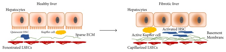
Liver fibrosis is a wound-healing response to chronic liver injury such as alcoholic/nonalcoholic fatty liver disease and viral hepatitis with no FDA-approved treatments. Liver fibrosis results in a continual accumulation of extracellular matrix (ECM) proteins and paves the way for replacement of parenchyma with nonfunctional scar tissue. The fibrotic condition results in drastic changes in the local mechanical, chemical, and biological microenvironment of the tissue. Liver parenchyma is supported by an efficient network of vasculature lined by liver sinusoidal endothelial cells (LSECs). These nonparenchymal cells are highly specialized resident endothelial cell type with characteristic morphological and functional features. Alterations in LSECs phenotype including lack of LSEC fenestration, capillarization, and formation of an organized basement membrane have been shown to precede fibrosis and promote hepatic stellate cell activation. Here, we review the interplay of LSECs with the dynamic changes in the fibrotic liver microenvironment such as matrix rigidity, altered ECM protein profile, and cell-cell interactions to provide insight into the pivotal changes in LSEC physiology and the extent to which it mediates the progression of liver fibrosis. Establishing the molecular aspects of LSECs in the light of fibrotic microenvironment is valuable towards development of novel therapeutic and diagnostic targets of liver fibrosis.
Conflict of interest statement
The authors declare that there is no conflict of interests regarding the publication of this paper.
Figures
References
Publication types
MeSH terms
Grants and funding
LinkOut - more resources
Full Text Sources
Other Literature Sources
Medical
