Review
. 2017 Mar 15;9(3):44.
doi: 10.3390/v9030044.

Reverse Transcription in the Saccharomyces cerevisiae Long-Terminal Repeat Retrotransposon Ty3

Affiliations
Review

Reverse Transcription in the Saccharomyces cerevisiae Long-Terminal Repeat Retrotransposon Ty3

Jason W Rausch et al. Viruses. .

Abstract

Converting the single-stranded retroviral RNA into integration-competent double-stranded DNA is achieved through a multi-step process mediated by the virus-coded reverse transcriptase (RT). With the exception that it is restricted to an intracellular life cycle, replication of the Saccharomyces cerevisiae long terminal repeat (LTR)-retrotransposon Ty3 genome is guided by equivalent events that, while generally similar, show many unique and subtle differences relative to the retroviral counterparts. Until only recently, our knowledge of RT structure and function was guided by a vast body of literature on the human immunodeficiency virus (HIV) enzyme. Although the recently-solved structure of Ty3 RT in the presence of an RNA/DNA hybrid adds little in terms of novelty to the mechanistic basis underlying DNA polymerase and ribonuclease H activity, it highlights quite remarkable topological differences between retroviral and LTR-retrotransposon RTs. The theme of overall similarity but distinct differences extends to the priming mechanisms used by Ty3 RT to initiate (-) and (+) strand DNA synthesis. The unique structural organization of the retrotransposon enzyme and interaction with its nucleic acid substrates, with emphasis on polypurine tract (PPT)-primed initiation of (+) strand synthesis, is the subject of this review.

Keywords: DNA polymerase; Ty3; retroelement; retrotransposon; reverse transcriptase; reverse transcription; ribonuclease H (RNase H).


Conflict of interest statement

The authors declare no conflict of interest. The funding sponsors had no role in the design of the study; in the collection, analyses, or interpretation of data; in the writing of the manuscript, and in the decision to publish the results.

Figures

Figure 1
Figure 1
Ty3 Reverse Transcription Cycle. (A) Structure of the double stranded preintegrative Ty3 DNA (black). U3, unique 3′ sequence; R, repeat sequence; U5, unique 5′ sequence; PBS, primer binding site; PPT, polypurine tract; (B) Genomic RNA is depicted in red. The bipartite nature of the PBS comprises sequences from both the 5′ PBS and the 3′ U3 regions; (C) Simplified initiation complex excluding the transfer RNA (tRNA) 5′ terminal nucleotides; (D) (−) strand strong stop synthesis, with concomitant degradation of genomic RNA by RNase H. Newly synthesized (−) strand DNA is shown in blue; (E) (−) strand transfer; (F) (−) strand synthesis and concomitant degradation of genomic RNA by RNase H; (G) (+) strand synthesis initiates from the PPT and extends into tRNA. Nascent (+) strand DNA is shown in green; (H) PPT is re-cleaved from (+) strand DNA and tRNA is cleaved from (−) strand DNA by RNase H; (I) Second (+) strand DNA, indicated in blue, displaces first; (J) PPT is again cleaved; (K) Third (+) strand synthesis initiates, and displaces second (+) strand; (L) Second (+) strand transfers to 3′-end of (−) DNA and PPT is cleaved; (M) Synthesis of both (+) and (−) strands is completed.
Figure 2
Figure 2
Structure of the asymmetric Ty3 RT homodimer in complex with its PPT-containing RNA/DNA hybrid. DNA and RNA strands of the cartoon representation are denoted in cyan and yellow, respectively. Subunit domains are color coded blue, red, green, and orange for fingers, palm, thumb, and RNase H, respectively, and the darker shading represents subunit A. Note the absence of a connection subdomain, a significant contrast between retroviral and LTR-retrotransposon RTs. Adapted from [37].
Figure 3
Figure 3
Contacts between Ty3 RT subunits A and B and the PPT-containing RNA/DNA hybrid. Color coding is consistent with subdomain designation of Figure 2, and DNA and RNA nucleotides are denoted in capital and small letters, respectively. The scissile PPT/U3 junction has been indicated, and base numbering is relative to substrate bound at the DNA polymerase active site Subunit B contacts are denoted “B” and circled. Parallel horizontal lines indicate van der Waals interactions. Diagonal and vertical lines indicate interactions mediated by the protein backbone (cyan) or side chains (black).
Figure 4
Figure 4
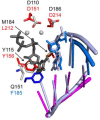
Alignment of the DNA polymerase active sites of Ty3 (PDB ID 4OL8, REF) and HIV-1 RT (PDB ID:1RTD). Carbon atoms of select Ty3 RT residues are shown in red (palm) and blue (fingers), and those of HIV-1 residues are in grey. The two catalytic metal ions and incoming dTTP are shown in grey and dark grey, respectively. Both HIV-1 DNA strands are shown as a light blue ladder, and the RNA template and DNA primer bound by Ty3 RT are shown in magenta and marine, respectively. The 3′-terminal nucleotides in both DNA primer strands are shown in stick form, and the stick radius of the incoming dTTP has been slightly expanded for contrast. Adapted from [37].
Figure 5
Figure 5
Phenotypic mixing strategy to determine the RNase H-competent Ty3 RT subunit. RNase H defective (D426N) and dimerization defective (R140A/R203A) mutant monomers are indicated in blue and grey, respectively. Notations d+ and d indicate a dimerization-competent and dimerization-incompetent subunit interface, while r+ and r denote RNase H-competent and RNase H-incompetent, respectively. Note that the d mutant only prevents dimerization when in the A subunit position. When purified mutants are mixed, RNase H activity is only recovered in a reconstituted dimer whose subunit B contributes to RNase H activity.
Figure 6
Figure 6
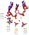
Alignment of RNase H active sites from Ty3 RT (PDB ID 4OL8, REF), Bacillus halodurans RNase H1 (PDB ID: 1ZB1, REF), and human RNase H1 (PDB ID: 2QK9, REF). Residue carbon atoms are shown in yellow, blue, and salmon, respectively. RNA strands from human and bacterial RNases H1 are shown in salmon and red, and two catalytic Mg++ ions from the Bh-RNase H1 structure are depicted as green spheres. The attacking nucleophilic water is shown as a red sphere.
Figure 7
Figure 7
(A) Model RNA/DNA hybrids to illustrate the specificity of cleavage at the Ty3 PPT/U3 junction. A hybrid containing the “all-RNA” strand, PPT/r, mimics selection of the PPT 3’-OH from the RNA/DNA replication mediate during (−) strand DNA synthesis, while a hybrid containing the RNA-DNA chimera, PPT/d, mimics release of the PPT 3’-OH from nascent DNA, an obligate step following initiation of (+) strand DNA synthesis; (B) experimental data. For both model substrates, the position of the PPT/U3 junction has been indicated. Adapted from [50].
Figure 8
Figure 8
Modulation of Ty3 PPT cleavage by targeted insertion of non-polar pyrimidine isosteres. (A) Representation of an A:T base pair and its A:F counterpart; (B) Model Ty3 RNA/DNA hybrid and a summary of pyrimidine isostere mutagenesis. DNA and RNA strands are depicted in capital and small letters, respectively, and the scissile PPT/U3 junction is indicated. Base-pair numbering is relative to the PPT/U3 junction (i.e., the last base of the PPT is denoted −1). Sites of cleavage relative to the position of T-F modification in the DNA strand are indicated; (C) experimental data. WT, unmodified hybrid, indicating cleavage at the PPT/U3 junction. For additional panels, the position of T-F modification in the DNA strand are indicated, and the asterisk illustrates the relocated RNase H cleavage in response to these modifications. Adapted from [50,51].

Similar articles

Cited by

References

    1. Telesnitsky A., Goff S.P. Reverse transcriptase and the generation of retroviral DNA. In: Coffin J.M., Hughes S.H., Varmus H.E., editors. Retroviruses. Cold Spring Harbor Laboratory Press; Cold Spring Harbor, NY, USA: 1997. pp. 121–160. - PubMed
    1. Baltimore D. RNA-dependent DNA polymerase in virions of RNA tumour viruses. Nature. 1970;226:1209–1211. doi: 10.1038/2261209a0. - DOI - PubMed
    1. Temin H.M., Mizutani S. RNA-dependent DNA polymerase in virions of rous sarcoma virus. Nature. 1970;226:1211–1213. doi: 10.1038/2261211a0. - DOI - PubMed
    1. Le Grice S.F. “In the beginning”: Initiation of minus strand DNA synthesis in retroviruses and LTR-containing retrotransposons. Biochemistry. 2003;42:14349–14355. doi: 10.1021/bi030201q. - DOI - PubMed
    1. Nowak E., Potrzebowski W., Konarev P.V., Rausch J.W., Bona M.K., Svergun D.I., Bujnicki J.M., Le Grice S.F., Nowotny M. Structural analysis of monomeric retroviral reverse transcriptase in complex with an RNA/DNA hybrid. Nucleic Acids Res. 2013;41:3874–3887. doi: 10.1093/nar/gkt053. - DOI - PMC - PubMed

MeSH terms

Substances

LinkOut - more resources