. 2017 May;58(5):858-871.
doi: 10.1111/epi.13719. Epub 2017 Mar 11.

Cav 1.3 channels play a crucial role in the formation of paroxysmal depolarization shifts in cultured hippocampal neurons

Affiliations

Cav 1.3 channels play a crucial role in the formation of paroxysmal depolarization shifts in cultured hippocampal neurons

Victoria Stiglbauer et al. Epilepsia. 2017 May.

Abstract

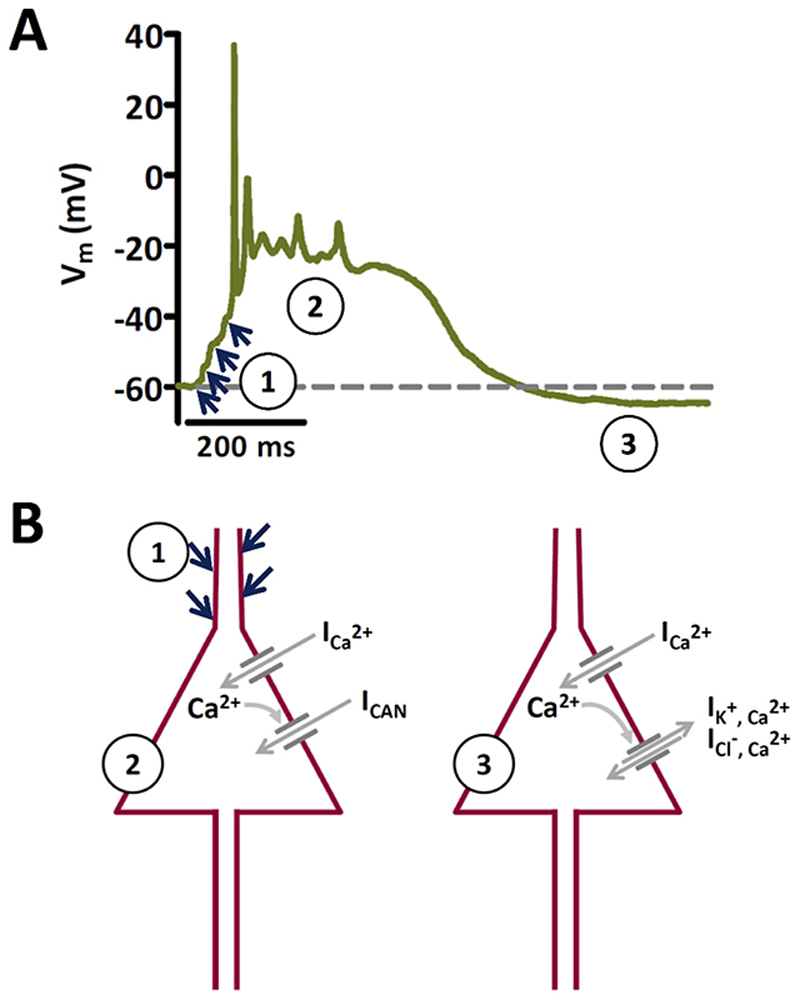
Objective: An increase of neuronal Cav 1.3 L-type calcium channels (LTCCs) has been observed in various animal models of epilepsy. However, LTCC inhibitors failed in clinical trials of epileptic treatment. There is compelling evidence that paroxysmal depolarization shifts (PDSs) involve Ca2+ influx through LTCCs. PDSs represent a hallmark of epileptiform activity. In recent years, a probable epileptogenic role for PDSs has been proposed. However, the implication of the two neuronal LTCC isoforms, Cav 1.2 and Cav 1.3, in PDSs remained unknown. Moreover, Ca2+ -dependent nonspecific cation (CAN) channels have also been suspected to contribute to PDSs. Nevertheless, direct experimental support of an important role of CAN channel activation in PDS formation is still lacking.

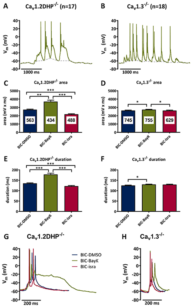
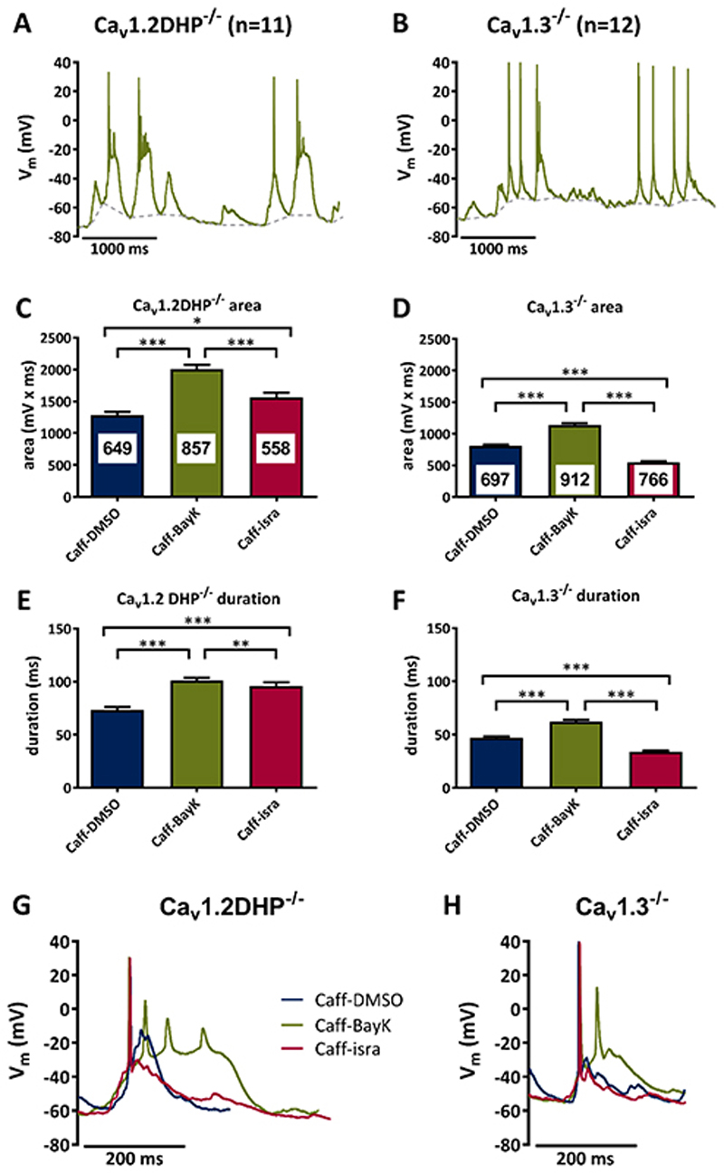
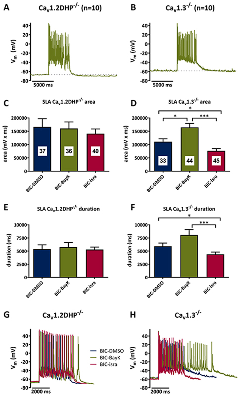
Methods: Primary neuronal networks derived from dissociated hippocampal neurons were generated from mice expressing a dihydropyridine-insensitive Cav 1.2 mutant (Cav 1.2DHP-/- mice) or from Cav 1.3-/- knockout mice. To investigate the role of Cav 1.2 and Cav 1.3, perforated patch-clamp recordings were made of epileptiform activity, which was elicited using either bicuculline or caffeine. LTCC activity was modulated using the dihydropyridines Bay K 8644 (agonist) and isradipine (antagonist).

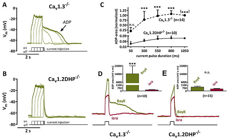
Results: Distinct PDS could be elicited upon LTCC potentiation in Cav 1.2DHP-/- neurons but not in Cav 1.3-/- neurons. In contrast, when bicuculline led to long-lasting, seizure-like discharge events rather than PDS, these were prolonged in Cav 1.3-/- neurons but not in Cav 1.2DHP-/- neurons. Because only the Cav 1.2 isoform is functionally coupled to CAN channels in primary hippocampal networks, PDS formation does not require CAN channel activity.

Significance: Our data suggest that the LTCC requirement of PDS relates primarily to Cav 1.3 channels rather than to Cav 1.2 channels and CAN channels in hippocampal neurons. Hence, Cav 1.3 may represent a new therapeutic target for suppression of PDS development. The proposed epileptogenic role of PDSs may allow for a prophylactic rather than the unsuccessful seizure suppressing application of LTCC inhibitors.

Keywords: CAN channels; Epileptogenesis; L-type voltage-gated calcium channels; Primary cultured hippocampal neurons.


Conflict of interest statement

Disclosure

None of the authors has any conflict of interest to disclose. We confirm that we have read the Journal’s position on issues involved in ethical publication and affirm that this report is consistent with those guidelines.

Figures

Figure 1
Figure 1. Proposed mechanisms of PDS generation.
A Recording of an original type of PDS that illustrates activation by summing up of excitatory inputs (indicated by the blue arrows) to a giant post-synaptic potential that triggers action potential firing (1), which turns after spike-decline into a plateau phase (2). After the plateau, the membrane potential may return back to baseline (dashed line) or transiently to an even more hyperpolarized potential (afterhyperpolarization) as in the example shown (3). B The two schemes illustrate the conductances that have been proposed to underlie the formation of original PDS. Phase 1 in A is mediated by excitatory synaptic input, presumably via AMPA type glutamate receptors. The decline in action potential amplitude is thought to be due to progressive inactivation of voltage-gated sodium channels. The plateau phase (2) may be largely due to L-type mediated Ca2+ inward current (ICa 2+), allegedly together with cationic inward current via CAN channels (ICAN). Afterhyperpolarizations (3) are considered to be due to K+-efflux and/or Cl--influx via Ca2+-activated K+ and Cl- channels. Note that this figure is based on a textbook figure published by Speckmann and Walden (1993) to illustrate the proposed role of CAN channels. It has been modified with respect to the prevailing view of an initiation of the PDS by excitatory postsynaptic potentials, , , whereas in the original depiction by Speckmann and colleagues the onset was described to be caused directly by inward calcium current.
Figure 2
Figure 2. Bicuculline-induced PDS require Cav1.3 channels.
A, B Suprathreshold electrical events recorded in Cav1.2DHP-/- neurons (A) or Cav1.3-/- neurons (B) after co-administration of bicuculline (10 μM) and BayK (3 μM). C-F Analysis of the area (C, D) and duration (E, F) of all suprathreshold electrical events recorded in the presence of bicuculline in Cav1.2DHP-/- neurons (C, E) or Cav1.3-/- neurons (D, F) under three conditions of LTCC activity: endogenous activity (DMSO control), potentiated (BayK, 3 μM) and inhibited (isradipine, 3 μM). The figures in the bars indicate the total number of events evaluated. All statistically significant differences are indicated by horizontal brackets. G, H Overlay of typical suprathreshold electric events recorded from Cav1.2DHP-/- neurons (G) or Cav1.3-/- neurons (H) from the experiments analysed in C to F.
Figure 3
Figure 3. Caffeine-induced PDS require Cav1.3 channels.
A, B Suprathreshold electrical events recorded in Cav1.2DHP-/- neurons (A) or Cav1.3-/- neurons (B) after co-administration of caffeine (1 mM) and BayK (3 μM). C-F Analysis of the area (C, D) and duration (E, F) of suprathreshold electrical events recorded in the presence of caffeine in Cav1.2DHP-/- neurons (C, E) or Cav1.3-/- neurons (D, F) under three conditions of LTCC activity: endogenous activity (DMSO control), potentiated (BayK, 3 μM) and inhibited (isradipine, 3 μM). The figures in the bars indicate the total number of events evaluated. All statistically significant differences are indicated by horizontal brackets. G, H Overlay of typical suprathreshold electric events recorded from Cav1.2DHP-/- neurons (G) or Cav1.3-/- neurons (H) from the experiments analysed in C to F.
Figure 4
Figure 4. Potentiation of Cav1.2 but not Cav1.3 channels gives rise to pronounced afterdepolarizations.
A, B Voltage response to brief current injections (50 to 1050 ms duration, increased in 4 steps of 250 ms) that cause depolarizations up to -20 mV in 0.5 μM tetrodotoxin-silenced neurons are shown together with their afterpotentials upon LTCC channel potentiation with 3 μM BayK. Distinct afterdepolarizations (ADP) can be seen in Cav1.3-/- neurons (A) but not in Cav1.2DHP-/- neurons (B). C Summary graph of experiments such as the one illustrated by original traces in A and B for ADP areas of 14 Cav1.3-/- neurons and of 10 Cav1.2DHP-/- neurons. Data (shown as mean + standard deviation) were normalized to the ADP area at 1050 ms current-injection duration, which is set to 1 for Cav1.3-/- neurons and 0.077 for Cav1.2DHP-/- neurons, which corresponds to the mean ADP area at this current-injection duration in relation to the mean ADP area of Cav1.3-/- neurons. n.s., not significant. (***) indicates that the statistical test had to be been done on the raw data rather than the normalized one for this data point. D, E Overlay of voltage responses to similar current injections in the presence of BayK and isradipine of Cav1.3-/- neurons and of Cav1.2DHP-/- neurons. Afterpotentials are blocked by 3 μM isradipine. Note that the y-axis shown in B also applies for the traces in D and E. The graphs in the inserts compare the maximum ADP areas (shown as mean + SEM) that were elicited in the presence of BayK or isradipine (isra) of Cav1.3-/- neurons (D, n= 10) and of Cav1.2DHP-/- neurons (E, n=15). n.s., not significant.
Figure 5
Figure 5. Implication of Cav1.2 and Cav1.3 channels in seizure-like activity.
A, B Examples of seizure-like activity (SLA) induced by co-application of bicuculline (10 μM) and BayK (3 μM) in Cav1.2DHP-/- neurons (A) or Cav1.3-/- neurons (B). C-F Analysis of the area (C, D) and duration (E, F) of SLA induced by 10 μM bicuculline in Cav1.2DHP-/- neurons (C, E) or Cav1.3-/- neurons (D, F) under three conditions of LTCC activity: endogenous activity (DMSO control), potentiated (BayK, 3 μM) and inhibited (isradipine, 3 μM). The figures in the bars indicate the total number of events evaluated. All statistically significant differences are indicated by horizontal brackets. G, H Overlay of typical SLA recorded from Cav1.2DHP-/- neurons (G) or Cav1.3-/- neurons (H) from the experiments analysed in C to F.

Similar articles

Cited by

References

    1. Calin-Jageman I, Lee A. Cav1 L-type Ca2+ channel signaling complexes in neurons. J Neurochem. 2008;105:573–583. - PubMed
    1. Zhang Z, Séguéla P. Metabotropic induction of persistent activity in layers II/III of anterior cingulate cortex. Cereb Cortex. 2010;20:2948–2957. - PubMed
    1. Pérez-Garci E, Larkum ME, Nevian T. Inhibition of dendritic Ca2+ spikes by GABAB receptors in cortical pyramidal neurons is mediated by a direct Gi/o-β-subunit interaction with Cav1 channels. J Physiol. 2013;591:1599–1612. - PMC - PubMed
    1. Kulak W, Sobaniec W, Wojtal K, et al. Calcium modulation in epilepsy. Pol J Pharmacol. 2004;56:29–41. - PubMed
    1. Amano H, Amano T, Matsubayashi H, et al. Enhanced calcium influx in hippocampal CA3 neurons of spontaneously epileptic rats. Epilepsia. 2001;42:345–350. - PubMed

Publication types

Substances