Comparative Study
. 2017 Jul 1;525(10):2343-2357.
doi: 10.1002/cne.24208. Epub 2017 Apr 21.

Anatomy of the lobula complex in the brain of the praying mantis compared to the lobula complexes of the locust and cockroach

Affiliations
Comparative Study

Anatomy of the lobula complex in the brain of the praying mantis compared to the lobula complexes of the locust and cockroach

Ronny Rosner et al. J Comp Neurol. .

Abstract

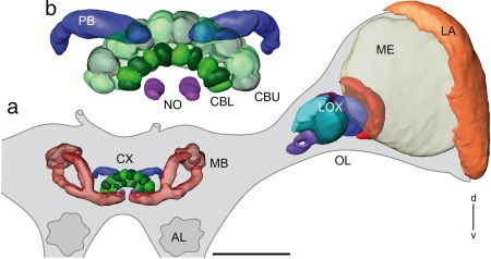
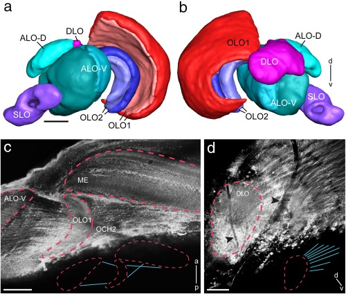
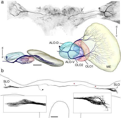
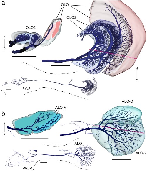
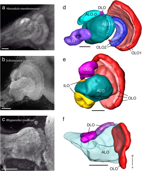
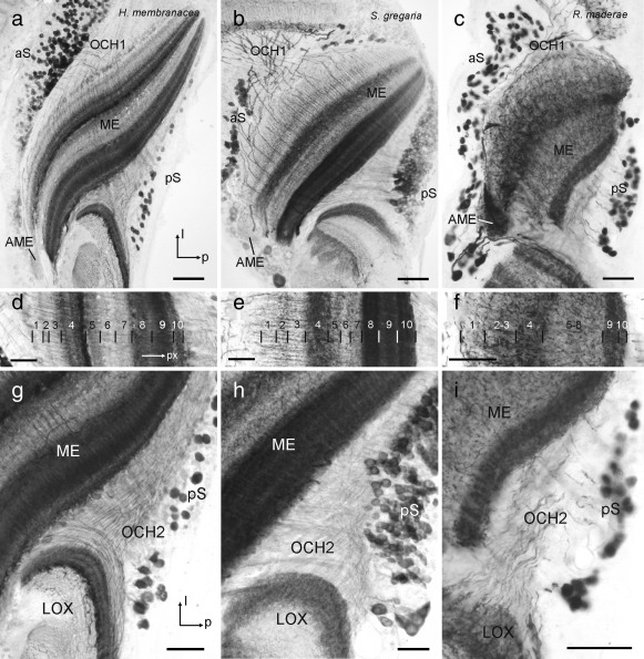
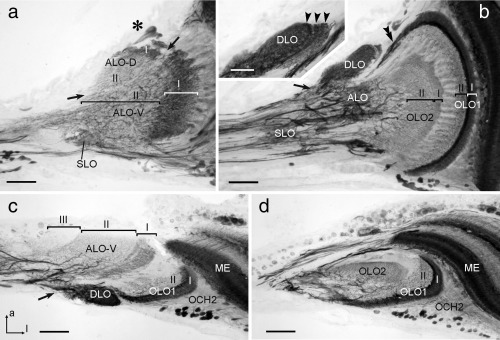
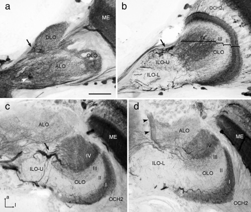
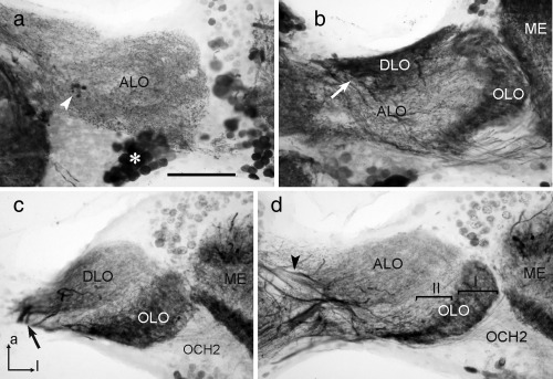
The praying mantis is an insect which relies on vision for capturing prey, avoiding being eaten and for spatial orientation. It is well known for its ability to use stereopsis for estimating the distance of objects. The neuronal substrate mediating visually driven behaviors, however, is not very well investigated. To provide a basis for future functional studies, we analyzed the anatomical organization of visual neuropils in the brain of the praying mantis Hierodula membranacea and provide supporting evidence from a second species, Rhombodera basalis, with particular focus on the lobula complex (LOX). Neuropils were three-dimensionally reconstructed from synapsin-immunostained whole mount brains. The neuropil organization and the pattern of γ-aminobutyric acid immunostaining of the medulla and LOX were compared between the praying mantis and two related polyneopteran species, the Madeira cockroach and the desert locust. The investigated visual neuropils of the praying mantis are highly structured. Unlike in most insects the LOX of the praying mantis consists of five nested neuropils with at least one neuropil not present in the cockroach or locust. Overall, the mantis LOX is more similar to the LOX of the locust than the more closely related cockroach suggesting that the sensory ecology plays a stronger role than the phylogenetic distance of the three species in structuring this center of visual information processing.

Keywords: RRID: AB_2313575; RRID: AB_2314457; RRID: AB_2315056; RRID: AB_2315425; RRID: AB_2336990; RRID: AB_2337244; RRID: AB_2338006; RRID: AB_2338713; RRID: AB_261363; RRID: nif-0000-00262; cockroach; insect visual system; lobula complex; locust; praying mantis.


Figures

Figure 1
Figure 1
Three‐dimensional reconstructions of principle neuropils in the protocerebrum of the praying mantis Hierodula membranacea. (a) Frontal view of reconstructed neuropils of the central brain and the left optic lobe embedded in the outlined mantis brain. (b) Enlarged view of the reconstructed central complex with central body (CB), PB, and paired noduli. AL = antennal lobe; CBL = lower division of the CB; CBU= upper division of the CB; CX = central complex; d = dorsal; LA = lamina; LOX = lobula complex; MB = mushroom body; ME = medulla; NO = noduli; OL = optic lobe; PB = protocerebral bridge; v = ventral. Scale bar = 500 µm
Figure 2
Figure 2
Detailed layout of the lobula complex (LOX) in Hierodula membranacea. (a, b) Three‐dimensional reconstruction of LOX neuropils as seen from frontal (a) and posterior (b). (c, d) Projection views of multiple confocal images for visualization of connectivity patterns of key optic lobe neuropils. (c) The OLO1 is connected with the medulla via the second optic chiasm. The ALO receives retinotopic input from the OLO1 via uncrossed fibers. Medulla, OLO1 and ALO are outlined by red dashed lines. Inset shows outline of connectivity pattern between medulla, OLO1 and ALO. (d) The DLO receives retinotopic from the medulla. The DLO is outlined by red dashed line. Black arrowheads point toward trachea present at the surface of the optic lobe. Inset shows outline of connectivity pattern between DLO and medulla. a = anterior; ALO‐D = dorsal subunit of the anterior lobe; ALO‐V = ventral subunit of the anterior lobe; d = dorsal; DLO = dorsal lobe; ME = medulla; OCH2 = second optic chiasm; OLO1 = outer lobe 1; OLO2 = outer lobe 2; p = posterior; SLO = stalk lobe; v = ventral; Scale bars = 100 µm (a–d)
Figure 3
Figure 3
Commissural neurons ramifying in the optic lobes of the praying mantis. (a, b) Neurons stained by injection of Neurobiotin via intracellular micropipettes. (a) Projection view of multiple confocal images of MELOXcom neuron in Rhombodera basalis. The medullae are truncated because of poor visibility in projection view and for reasons of space. Below: Frontal and ventral views of three‐dimensional reconstruction of optic lobe neuropils and neuron ramifications in the ipsilateral medulla and LOX. (b) Commissural neuron (SLOcom) ramifying in the stalk lobes (SLO) of both optic lobes and in the dorsal protocerebrum. The sites of arborizations in the dorsal protocerebrum are indicated by red arrowheads. The image of the neuron was generated as projection view from confocal image stack by masking background. Insets show smooth ramifications in the SLO ipsilateral to the cell's soma and beaded endings in the contralateral SLO. a = anterior; ALO‐D = dorsal compartment of the anterior lobe; ALO‐V = ventral compartment of the anterior lobe; d = dorsal; ME = medulla; OLO1 = outer lobe 1; OLO2 = outer lobe 2; p = posterior; v = ventral. Scale bars = 100 µm (a and b)
Figure 4
Figure 4
Three‐dimensional reconstructions of tangential neurons ramifying in the LOX of Hierodula membranacea. (a, b) Two projection neurons were stained during intracellular recordings. Both neurons have ramifications in the posterior ventrolateral protocerebrum (insets) and the LOX. (a) Projection neuron from the outer lobe, termed TOpro1‐neuron. Extensive dendritic arborizations are confined to the two layers of OLO2 and the most proximal layer of OLO1 as is especially apparent in horizontal profile view. (b) Frontal view and horizontal section showing dendritic tree of projection neuron termed TApro1‐neuron with ramifications in two layers of the ALO‐V and in the ALO‐D. Red dotted lines in (a) and (b) indicate planes of horizontal sections. a = anterior; ALO = anterior lobe; OLO1 = outer lobe 1; OLO2 = outer lobe 2; p = posterior; PVLP = posterior ventrolateral protocerebrum. Scale bars = 200 µm (a, b)
Figure 5
Figure 5
Lobula complexes (LOXs) of the praying mantis Hierodula membranacea, the locust Schistocerca gregaria, and the cockroach Rhyparobia maderae. (a–c) Projection views of synapsin immunostained LOXs of the praying mantis (a), locust (b), and cockroach (c) obtained from confocal image stacks. (d–f) Three‐dimensional reconstructions of the LOXs shown in (a–c). ALO = anterior lobe; ALO/ILO = anterior lobe inner lobe aggregate; d = dorsal; DLO = dorsal lobe; ILO = inner lobe; OLO = outer lobe; OLO1 = outer lobe 1; OLO2 = outer lobe 2; SLO = stalk lobe; v = ventral. Scale bars = 100 µm (a–f)
Figure 6
Figure 6
γ‐Aminobutyric acid (GABA) immunostaining in the medulla (ME) and second optic chiasm (OCH2) of the praying mantis H. membranacea (a, d, g), the locust S. gregaria (b, e, h) and the cockroach R. maderae (c, f, i). (a–c) Horizontal sections through the medulla at the level of the accessory medulla (AME). In all three species immunostained somata are scattered in the anterior soma rind (aS) near the first optic chiasm (OCH1) and in the posterior soma rind (pS) adjacent to OCH2. The AME is virtually free of immunostaining in the mantis (a) and locust (b), but shows dense immunostaining in the cockroach (c). l = lateral; p = posterior. (d–f) Horizontal sections through the medulla illustrating the distribution of immunostaining across medulla layers. The ten layers (1–10 from distal to proximal) in the ME of S. gregaria conform to previous studies (e.g., Beetz et al., 2015). Layering in H. membranacea (d) and R. maderae was adjusted to the layering scheme of S. gregaria for easier comparison. px = proximal. (g–i) Horizontal sections illustrating fiber trajectories in the second optic chiasm (OCH2) of the three species. Scale bars = 100 µm in (a, b); 50 µm in (c, d–i)
Figure 7
Figure 7
γ‐Aminobutyric acid (GABA) immunostaining in the lobula complex (LOX) of the praying mantis H. membranacea. (a, b) Frontal sections at an anterior (a) and an intermediate (b) level through the LOX. (c, d) Horizontal sections at a dorsal (c) and ventral (d) level. Sections illustrate immunostaining in the anterior lobe (ALO‐D, ALO‐V), dorsal lobe (DLO), outer lobe 1 and 2 (OLO1, OLO2), and the stalk lobe (SLO). Inset in (b) illustrates layered organization of the DLO (arrowheads). The anterior lobe can be subdivided into a dorsal and a ventral unit (ALO‐D, ALO‐V, divide indicated by arrows in (a). Based on distinct differences in immunostaining, 2 layers can be distinguished in the ALO‐D, OLO1, and OLO2 (labeled I and II, respectively), and three layers in the ALO‐V (labeled I–III). Asterisk in (a) indicates immunoreactive somata of tangential neurons near the dorsal face of the ALO‐D. Arrows in (b) (except inset) and (c) point to immunostained fibers entering the DLO. Double arrowhead in (a) points to immunoreactive fibers connecting OLO1 with the central brain. OLO1 is, in addition, innervated by columnar neurons from the medulla (ME) with fibers passing through the second optic chiasm (OCH2). a = anterior; l = lateral (applies to c and d). Scale bars = 200 µm; 50 µm in inset of (a)
Figure 8
Figure 8
γ‐Aminobutyric acid (GABA) immunostaining in the lobula complex (LOX) of the locust S. gregaria. (a, b) Frontal sections at an anterior (a) and a more posterior (b) level; (c, d) horizontal sections at a dorsal (c) and ventral (d) level of the LOX. Distinct immunostaining is present in the anterior lobe (ALO), dorsal lobe (DLO), inner lobe (ILO‐U, ILO‐L), and outer lobe (OLO) of the LOX. In the OLO, four major layers (labeled I–IV) can be distinguished based on different density of GABA immunostaining. The ILO can be further subdivided into an upper half (ILO‐U) with sparse varicose immunostained processes and a lower half (ILO‐L) which is nearly devoid of immunostaining. Arrows in (a, b, and c) point to immunostained fibers entering the DLO and layer IV of the OLO. Arrowheads in (a) and (d) indicate a prominent fiber bundle connecting the ALO with the central brain via the anterior optic tract. Immmunostained fibers in the second optic chiasm (OCH2) connect the medulla (ME) to the OLO. a = anterior; l = lateral (applies to c and d). Scale bar = 100 µm (applies to a–d)
Figure 9
Figure 9
γ‐Aminobutyric acid (GABA) immunostaining in the lobula complex (LOX) of the cockroach R. maderae. (a, b) Frontal sections at an anterior (a) and a more posterior (b) level; (c, d) horizontal sections at a dorsal (c) and ventral (d) level of the LOX. Based on distinct GABA immunostaining, an anterior/inner lobe (ALO/ILO), a dorsal lobe (DLO), and an outer lobe (OLO) can be distinguished. In the OLO, a strongly stained distal layer (I) and a sparsely innervated second proximal layer (II) can be distinguished (d). Arrows in (b) and (c) point to immunostained fibers entering the DLO. Arrowheads in (a) and (d) point to a bundle of immunoreactive fibers connecting the central brain and ALO via the anterior optic tract. Asterisk in (a) points to cell bodies whose primary neurites project to the central brain. Immunostained fibers in the second optic chiasm (OCH2) connect the medulla (ME) to the OLO. a = anterior, l = lateral (applies to c and d). Scale bar = 100 µm (applies to a–d)

Similar articles

Cited by

References

    1. Aptekar, J. W. , Keleş, M. F. , Lu, P. M. , Zolotova, N. M. , & Frye, M. A. (2015). Neurons forming optic glomeruli compute figure–ground discriminations in Drosophila . The Journal of Neuroscience: The Official Journal of the Society for Neuroscience, 35, 7587–7599. - PMC - PubMed
    1. Beetz, M. J. , el Jundi, B. , Heinze, S. , & Homberg, U. (2015). Topographic organization and possible function of the posterior optic tubercles in the brain of the desert locust Schistocerca gregaria . The Journal of Comparative Neurology, 523, 1589–1607. - PubMed
    1. Berger, F. A. (1985). Morphologie und Physiologie einiger visueller Interneuronen in den optischen Ganglien der Gottesanbeterin Mantis religiosa. (Doctoral dissertation). University of Düsseldorf, Düsseldorf, Germany.
    1. Borst, A. , Haag, J. , & Reiff, D. F. (2010). Fly motion vision. Annual Review of Neuroscience, 33, 49–70. - PubMed
    1. Borst, A. , & Helmstaedter, M. (2014). Common circuit design in fly and mammalian motion vision. Nature Neuroscience, 18, 1067–1076. - PubMed

Publication types

LinkOut - more resources