Connectomics of synaptic microcircuits: lessons from the outer retina
- PMID: 28295344
- PMCID: PMC5556146
- DOI: 10.1113/JP273671
Connectomics of synaptic microcircuits: lessons from the outer retina
Abstract
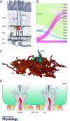
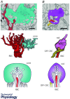
Photoreceptors form a sophisticated synaptic complex with bipolar and horizontal cells, transmitting the signals generated by the phototransduction cascade to downstream retinal circuitry. The cone photoreceptor synapse shows several characteristic anatomical connectivity motifs that shape signal transfer: typically, ON-cone bipolar cells receive photoreceptor input through invaginating synapses; OFF-cone bipolar cells form basal synapses with photoreceptors. Both ON- and OFF-cone bipolar cells are believed to sample from all cone photoreceptors within their dendritic span. Electron microscopy and immunolabelling studies have established the robustness of these motifs, but have been limited by trade-offs in sample size and spatial resolution, respectively, constraining precise quantitative investigation to a few individual cells. 3D-serial electron microscopy overcomes these limitations and has permitted complete sets of neurons to be reconstructed over a comparatively large section of retinal tissue. Although the published mouse dataset lacks labels for synaptic structures, the characteristic anatomical motifs at the photoreceptor synapse can be exploited to identify putative synaptic contacts, which has enabled the development of a quantitative description of outer retinal connectivity. This revealed unexpected exceptions to classical motifs, including substantial interaction between rod and cone pathways at the photoreceptor synapse, sparse photoreceptor sampling and atypical contacts. Here, we summarize what was learned from this study in a more general context: we consider both the implications and limitations of the study and identify promising avenues for future research.
Keywords: bipolar cell; connectomics; electron microscopy; photoreceptor; retina; synapse.
© 2017 The Authors. The Journal of Physiology © 2017 The Physiological Society.
Figures


References
-
- Baden T, Schubert T, Chang L, Wei T, Zaichuk M, Wissinger B & Euler T (2013). A tale of two retinal domains: Near‐optimal sampling of achromatic contrasts in natural scenes through asymmetric photoreceptor distribution. Neuron 80, 1206–1217. - PubMed
Publication types
MeSH terms
LinkOut - more resources
Full Text Sources
Other Literature Sources
Miscellaneous

